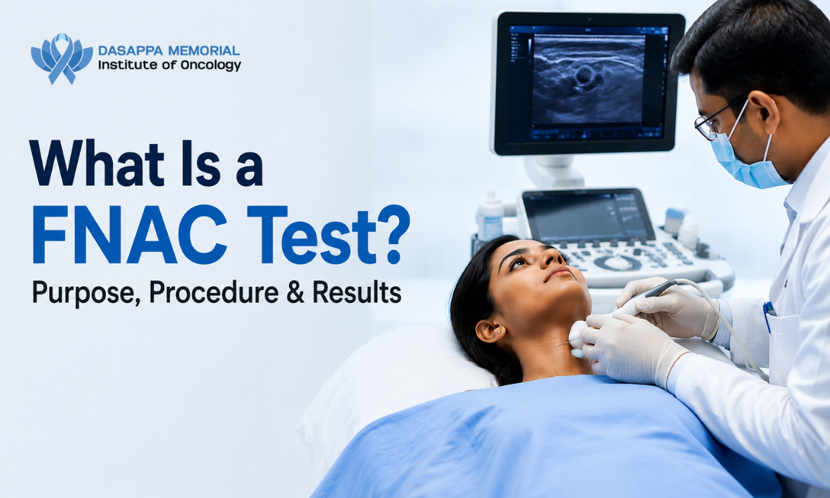
Discovering an unexplained lump in your neck, thyroid, or breast can trigger immediate anxiety, but getting clear answers early is key to regaining peace of mind. When clinicians evaluate suspicious tissue swellings, understanding what the FNAC test (Fine Needle Aspiration Cytology) is helps demystify this quick, minimally invasive diagnostic investigation. As a routine first-line assessment at the Best cancer hospital in Bangalore, an FNAC uses an ultra-thin needle to collect microscopic cell samples directly from tissue masses.
This safe outpatient procedure allows expert cytopathologists to quickly distinguish benign fluid cysts from cellular conditions that require specialized medical attention.
Unlike a core needle or surgical open biopsy, which removes intact slices of tissue architecture using larger hollow instruments or surgical incisions, a Fine Needle Aspiration Cytology focuses directly on individual cellular clusters.
The needle utilized is often thinner than the standard gauge used during a routine arm vein blood draw. By harvesting loose cells from suspicious nodules, doctors can analyze cellular structures under a microscope without causing surgical trauma or requiring downtime for recovery.
Our core philosophy at Dasappa Cancer Hospital of “healing with heart” means framing diagnostic steps as empowering milestones rather than intimidating procedures. We pair high-resolution real-time ultrasound guidance with immediate on-site cytological evaluations, ensuring that every patient receives rapid, highly accurate answers in a supportive and comforting environment.
Medical Disclaimer
The diagnostic protocols, procedural descriptions, and cytopathological classification frameworks outlined in this guide are intended strictly for educational and public health awareness purposes. This content does not constitute formal medical advice, clinical diagnosis, or a personalized treatment plan. Fine needle aspiration requirements and head/neck evaluations vary significantly based on individual medical histories and physical presentations. Always consult a qualified physician, endocrinologist, or surgical oncologist to evaluate specific lumps, neck swellings, or diagnostic lab reports. Never disregard or delay seeking professional clinical care based on the general information provided in this article.
Purpose & Clinical Intent: When and Why FNAC Is Recommended
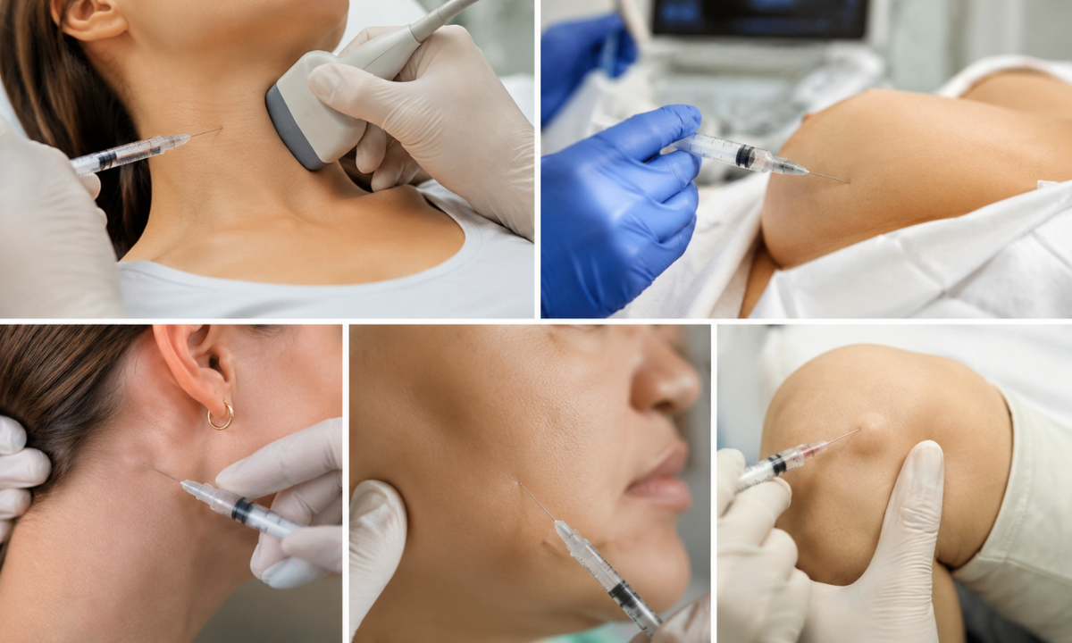
When a physician discovers an unexplained swelling, lump, or focal nodule during a routine physical examination or on an ultrasound scan, time is of the essence to determine its cause. Understanding why the FNAC test is done gives patients confidence when their doctor orders this investigation, as it provides a rapid, reliable, and non-surgical first assessment of suspicious tissue changes.
Instead of subjecting a patient to invasive surgical exploratory procedures, fine needle cytology acts as a diagnostic bridge distinguishing simple, fluid-filled benign cysts and inflammatory reactions from conditions requiring structured oncological care.
Key Clinical Indications & Anatomical Sites
Fine needle aspiration is versatile and can evaluate tissue masses across various superficial and deep anatomical regions:
| Targeted Body Region | Clinical Presentation | Primary Diagnostic Objective |
|---|---|---|
| Thyroid Gland | Single or multiple nodules, enlarged goiters, or sudden anterior neck swellings. | Differentiates benign multinodular goiters or thyroiditis from papillary, follicular, or medullary thyroid carcinomas. |
| Cervical Lymph Nodes | Persistent, enlarged, or hardened lymph nodes in the neck, under armpits, or groin. | Determines if swelling is due to reactive infection (e.g., tuberculosis) or metastatic spread from head and neck tumors. |
| Salivary Glands | Swellings in the parotid, submandibular, or sublingual glands near the jawline. | Identifies benign pleomorphic adenomas, Warthin tumors, or malignant salivary gland lesions. |
| Breast & Axilla | Solitary palpable breast masses, dense fibrocystic changes, or suspicious axillary node enlargement. | Distinguishes harmless fibroadenomas and fluid cysts from early-stage breast carcinomas. |
| Soft Tissue & Skin | Subcutaneous nodules, deep tissue masses, or recurrent lesion sites. | Rules out soft tissue sarcomas, lipomas, or localized inflammatory reactions. |
Differentiating Benign Conditions from Malignancy
The primary goal of FNAC is to categorize suspicious tissue without causing undue physical stress or surgical delay:
- Ruling Out Infection & Inflammation: In regions like South Asia, enlarged lymph nodes are frequently caused by granulomatous infections such as tuberculosis or reactive lymphadenopathy from viral bugs. FNAC quickly identifies these benign, treatable conditions, sparing the patient unnecessary cancer scares.
- Preventing Unnecessary Surgery: If fine needle cytology reveals a simple fluid cyst or benign adenoma, patients can often avoid surgery entirely and opt for simple drainage or routine observation.
- Guiding Surgical & Oncological Planning: When malignant cells are identified, cytology provides the essential baseline data needed by surgical oncologists to plan definitive core biopsies, molecular testing, or direct minimally invasive surgery.
At Dasappa Cancer Hospital, our diagnostic team evaluates every sample with extreme care. By combining immediate cytological review with supportive patient communication, we ensure you receive fast, definitive diagnostic answers so you can take the next steps in your care with complete confidence.
The FNAC Journey Step-by-Step: What to Expect
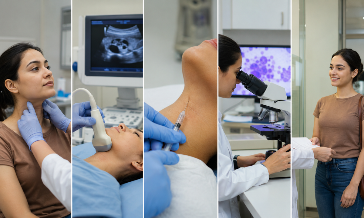
Understanding what happens during a fine needle aspiration can significantly ease pre-test anxiety. The clinical FNAC procedure is an efficient, virtually painless outpatient investigation that usually takes less than 15 to 20 minutes from setup to completion, requiring no general anesthesia or hospital admission.
At Dasappa Cancer Hospital, we prioritize patient comfort at every stage, combining high-resolution imaging with expert needle precision to make the sampling process as swift and gentle as possible.
The Step-by-Step Diagnostic Experience
- Patient Positioning & Local Preparation (3 to 5 Minutes): You will be comfortably seated or laid flat on an examination couch, depending on the site of the lump. The skin overlying the target area is thoroughly cleansed with an antiseptic solution. While the needle used is extremely fine, a mild topical numbing spray or small local anesthetic injection may be applied for sensitive areas to eliminate any discomfort.
- Ultrasound-Guided Needle Insertion (2 to 3 Minutes): To ensure absolute accuracy especially for deep, small, or sub-centimeter nodules our cytopathologist or radiologist uses real-time high-frequency ultrasound. This allows the specialist to visualize the needle’s tip in real time as it enters the target mass, avoiding surrounding blood vessels or delicate nerve structures.
- Cellular Aspiration & Sampling (Less than 60 Seconds): Using a fine-gauge needle (typically 22 to 25 gauge) attached to a syringe, the specialist performs gentle back-and-forth passes inside the nodule under controlled vacuum pressure. This movement collects loose cellular clusters inside the needle shaft without damaging surrounding tissue architecture.
- Rapid On-Site Evaluation (ROSE) (Immediate): Before you leave the room, the collected cells are smeared onto glass slides and stained immediately for a preliminary microscopic check. This Rapid On-Site Evaluation confirms that a sufficient quantity of high-quality cells was harvested, drastically reducing the chance of an inconclusive result or the need for a repeat visit.
- Post-Procedure Care & Discharge (2 to 3 Minutes): Once adequate samples are confirmed, the needle is withdrawn, and gentle pressure is applied to the site with a clean bandage to prevent minor bruising.
You can immediately return to your normal daily activities, with no restrictions on driving, walking, or working.
Managing Post-Procedure Comfort
As fine needle aspiration is minimally invasive, recovery is fast and uncomplicated:
- Minimal Soreness: Mild tenderness or a minor pinpoint bruise at the needle entry site is completely normal and typically resolves within 24 to 48 hours.
- Simple Relief: Standard over-the-counter pain relievers or a brief cold compress are usually more than sufficient if minor aching occurs.
You do not need to take time off work or arrange for overnight post-procedure monitoring, making FNAC one of the most patient-friendly diagnostic tools available in modern oncology.
Specialized Applications With Focus on Thyroid & Neck Swellings
The head and neck region contains a dense network of delicate anatomical structures, including major blood vessels, vital nerves, salivary glands, and the thyroid gland. As even minor swellings in this area can cause significant anxiety, specialized applications of fine needle aspiration require high precision and standardized clinical reporting.
When evaluating anterior neck masses, requesting a targeted FNAC test thyroid evaluation allows cytopathologists to categorize suspicious nodules accurately using international reporting standards, sparing most patients from unnecessary neck surgery while ensuring those with true malignancies receive prompt, definitive care.
The Bethesda System for Reporting Thyroid Cytopathology
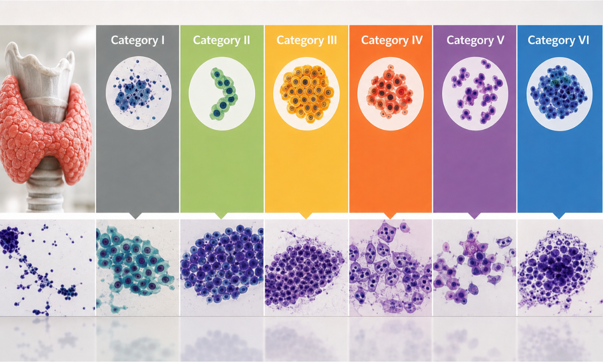
To eliminate diagnostic confusion between endocrinologists, surgeons, and pathologists, thyroid cytology reports follow the standardized Bethesda System (Categories I through VI). This classification directly links cellular findings to clinical risk and recommended next steps:
| Bethesda Category | Diagnostic Classification | Malignancy Risk | Standard Clinical Action |
|---|---|---|---|
| Category I | Non-Diagnostic / Unsatisfactory | 5 to 10% | Repeat ultrasound-guided aspiration after 6 to 8 weeks. |
| Category II | Benign (Colloid nodule, Thyroiditis) | 0 to 3% | Clinical observation & periodic ultrasound follow-up. |
| Category III | Atypia of Undetermined Significance (AUS) | 10 to 30% | Repeat FNAC, molecular testing, or selective diagnostic lobectomy. |
| Category IV | Follicular Neoplasm / Suspicious for FN | 15 to 40% | Surgical lobectomy (removal of the affected thyroid lobe). |
| Category V | Suspicious for Malignancy | 60 to 80% | Near-total or total thyroidectomy. |
| Category VI | Malignant (e.g., Papillary Thyroid Carcinoma) | 97 to 99% | Definitive surgical resection and oncological management. |
Cervical Lymph Nodes & Salivary Gland Evaluations
Beyond the thyroid gland, fine needle cytology plays a pivotal role in diagnosing swellings across other key structures in the neck:
- Cervical Lymph Node Staging: The neck contains over 300 lymph nodes. When a node remains persistently enlarged, FNAC quickly determines whether the swelling stems from a benign granulomatous infection (such as tuberculosis) or indicates metastatic spread from primary head, neck, or thoracic lesions.
- Salivary Gland Masses: Swellings in the parotid or submandibular glands are evaluated to differentiate benign tumors (like pleomorphic adenomas or Warthin tumors) from aggressive salivary carcinomas, giving surgical oncologists the exact map needed to protect the facial nerve during intervention.
Why Real-Time Ultrasound Guidance Matters: The anterior neck is crowded with major blood vessels like the carotid artery and jugular vein. Performing fine needle aspiration under continuous, real-time ultrasound imaging ensures the needle enters only the target nodule, completely avoiding adjacent vascular structures and maximizing cell yield.
By applying these standardized diagnostic frameworks, the team at Dasappa Cancer Hospital provides patients with complete clarity regarding their neck or thyroid swellings ensuring that every decision is backed by objective cellular data and compassionate expert guidance.
Decoding the Cytology Findings: Understanding Your Results
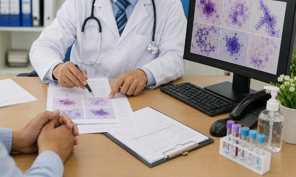
Receiving your lab results can feel overwhelming, especially if you are unfamiliar with medical terminology. Once your tissue sample is evaluated under a high-power microscope, a detailed diagnostic summary is generated. Reviewing your final FNAC report with your specialist helps translate complex microscopic cellular findings into a clear, actionable treatment plan.
A comprehensive cytopathology document does not simply state “yes” or “no.” It breaks down the specimen’s physical features, background components (such as blood, colloid, or fluid), and overall cellular arrangements.
Key Diagnostic Categories Explained
Cytopathologists group microscopic findings into four primary diagnostic categories:
1. Benign (Negative for Malignancy):
What it means: No cancerous or precancerous cells were identified in the sample. The mass is caused by a non-cancerous condition, such as a fluid cyst, colloid nodule, lipoma, or reactive inflammatory swelling.
Next Steps: Routine clinical follow-up or simple symptomatic treatment. No aggressive intervention is required.
2. Atypical / Suspicious (Equivocal):
What it means: The cells display unusual features or architectural patterns that differ from normal tissue, but the changes are not definitive enough to confirm a cancer diagnosis.
Next Steps: Your physician may recommend advanced ancillary testing (such as cell block preparation or immunohistochemistry), a repeat needle test, or a core needle biopsy to gain structural clarity.
3. Malignant (Positive for Malignancy):
What it means: Clear, unambiguous cancerous cells were detected in the microscopic field. The pathologist will often classify the specific cancer type (e.g., carcinoma, lymphoma, or melanoma).
Next Steps: Immediate consultation with a surgical oncologist or multidisciplinary cancer team to plan targeted staging and treatment strategies.
4. Inadequate / Non-Diagnostic:
What it means: The sample contained too few cells, excessive blood, or fluid artifacts to allow for a safe, reliable microscopic reading.
Next Steps: Repeating the sampling procedure ideally under real-time ultrasound guidance to ensure an adequate cellular yield.
What Happens If Results Are Inconclusive?
Inconclusive findings do not automatically mean bad news; they simply mean the needle collected insufficient cellular material for a definitive answer. At modern diagnostic centers, advanced techniques are used to minimize inconclusive outcomes:
- Cell Block & Biomarker Testing: Excess aspirated material can be processed into a paraffin wax block, allowing pathologists to perform specialized antibody stains (immunohistochemistry) to pinpoint cell origin.
- Ultrasound Re-Evaluation: If a mass is calcified, cystic, or very small, real-time imaging ensures the second pass precisely targets the solid, cell-rich portion of the nodule.
Our cytopathology specialists carefully review every finding alongside your physical examination data and imaging scans. This thorough approach ensures you receive an accurate diagnosis, eliminating guesswork and giving you a clear roadmap forward.
Multidisciplinary Excellence & Head/Neck Care at Dasappa
Discovering a suspicious lump in the neck, thyroid, or jawline requires more than just a standalone laboratory reading; it demands immediate, integrated clinical attention. As a recognized center of excellence for head & neck cancer treatment in Bangalore, Dasappa Cancer Hospital brings together diagnostic cytopathology, advanced imaging, and surgical oncology under a single roof to deliver fast, seamless care.
When facing potential head and neck malignancies, time and precision are paramount. Our diagnostic and surgical framework ensures that every patient moves from initial uncertainty to a clear, personalized treatment plan without unnecessary delays:
- Veteran Surgical Leadership: Led by Dr Pandu Dasappa, Chief Surgical Oncologist, our head and neck surgical teams possess decades of specialized experience in managing complex thyroid, parotid, oral, and cervical lymph node malignancies.
- Rapid On-Site Cytology Integration: By performing Fine Needle Aspiration Cytology with immediate on-site slide checks (ROSE), we ensure adequate cellular sampling on the very first visit, avoiding the frustration of repeat procedures.
- Nerve-Sparing Surgical Precision: For conditions requiring surgical resection such as thyroidectomies or parotidectomies, our surgeons utilize state-of-the-art intraoperative monitoring to protect delicate motor nerves, preserving facial expression and vocal function.
Beyond clinical and surgical precision, we provide dedicated psychosocial support, nutritional guidance, and speech/swallow rehabilitation, ensuring that both the physical and emotional well-being of your family are fully supported throughout the treatment journey.
Conclusion
A fine needle aspiration is far more than a routine diagnostic test; it is a fast, safe, and virtually painless key to unlocking peace of mind. Whether evaluating a silent thyroid nodule, an enlarged lymph node, or a persistent neck swelling, taking proactive steps early empowers you and your medical team to make informed, data-backed decisions. At Dasappa Cancer Hospital, we blend cutting-edge diagnostic technology with deep human compassion, ensuring that every patient receives the precise care, clarity, and support needed to walk the path toward complete recovery with confidence.