



Brain Tumor Symptoms That Should Never Be Ignored

Table of Contents

Recognizing early, subtle brain tumor symptoms is critical for preventing irreversible neurological damage and ensuring favorable clinical outcomes. As the human skull is a rigid, non-expandable container, any abnormal intracranial growth disrupts delicate pressure dynamics and impairs neurological function.

When persistent headaches, unprovoked seizures, or cognitive changes arise, seeking specialized evaluation for Brain Tumor Treatment in Bangalore connects patients with advanced neuro-diagnostic imaging, multidisciplinary tumor boards, and modern surgical onco-resection protocols designed to alleviate pressure and preserve vital brain function.

While everyday tension headaches and occasional fatigue are common responses to modern lifestyle stress, persistent neurological changes require careful clinical evaluation.

The fundamental reason brain tumors produce distinct, progressive symptoms lies in neuro-anatomy and intracranial physiology, governed by the Monro-Kellie Doctrine:

  • Fixed Intracranial Volume: The rigid adult cranium has a fixed internal volume housing three primary components: brain parenchyma (~80%), circulating cerebral blood (~10%), and cerebrospinal fluid (CSF, ~10%).
  • Loss of Compliance: In the early stages of tumor growth, the brain compensates by displacing CSF into the spinal subarachnoid space and reducing venous blood volume. Once these compensatory mechanisms are exhausted, even a tiny increase in tumor mass or surrounding swelling (peritumoral vasogenic edema) causes a steep, exponential rise in Intracranial Pressure (ICP).
  • Decompensation & Herniation Risks: Uncontrolled elevated ICP compromises cerebral perfusion, starves neurons of oxygen, and risks mechanical brain herniation across internal fibrous partitions (such as the falx cerebri or tentorium cerebelli).

What You Will Discover in This Guide

  • Anatomical Localization: How the exact anatomical subsite (frontal, temporal, parietal, occipital, cerebellar, or brainstem) determines your specific functional symptoms.
  • The “Never-Ignore” Emergency Checklist: Recognizing the classic signs of acute intracranial hypertension, new-onset adult seizures, and papilledema.
  • The Precision Diagnostic Cascade: Navigating advanced multiparametric contrast MRI, MR spectroscopy, functional mapping, and molecular tumor profiling.
  • Modern Multimodal Interventions: Understanding maximal safe neurosurgical resection, stereotactic radiosurgery, and targeted therapies.

Managing intracranial space-occupying lesions demands rapid diagnostic clarity, microsurgical precision, and integrated oncology expertise.

Led by Dr. Pandu Dasappa (M.Ch Surgical Oncology, GCRI Ahmedabad; with over 27 years of surgical oncology experience), Dasappa Cancer Hospital delivers evidence-based multidisciplinary care, ensuring patients receive accurate staging, surgical intervention, and personalized adjuvant therapy protocols.

Medical Disclaimer

The clinical descriptions, anatomical localization models, diagnostic pathways, and treatment overviews presented in this article are provided strictly for educational and public health awareness purposes. This content does not constitute formal medical advice, clinical diagnosis, or a personalized neurosurgical treatment plan. Any new-onset seizures, persistent headaches, visual changes, or suspected intracranial abnormalities must be evaluated promptly by a qualified neurosurgeon, surgical oncologist, medical oncologist, or licensed medical professional.

Anatomical Localization: How Tumor Location Dictates Functional Deficits

The brain is an intricately mapped organ where specific anatomical regions govern distinct cognitive, sensory, and motor functions. As brain parenchyma itself lacks pain receptors, a space-occupying lesion often produces subtle, localized neurological symptoms long before it elevates global intracranial pressure.

Recognizing these focal deficits allows clinicians to pinpoint the precise anatomical lobe or subsite compromised by tumor expansion or peritumoral edema.

Anatomical Functional Deficit Matrix

Cerebral / Infratentorial RegionPrimary Physiological FunctionHallmark Focal Deficits 
Frontal LobeExecutive function, motor planning, emotional regulation, and expressive speech (Broca’s area).• Progressive personality changes, apathy, or uncharacteristic disinhibition• Contralateral motor weakness or hemiparesis• Expressive aphasia (struggling to speak despite knowing what to say)• Impaired judgment and loss of workplace problem-solving skills
Temporal LobeAuditory processing, long-term memory retrieval, and language comprehension (Wernicke’s area).• Receptive aphasia (fluent but nonsensical speech, difficulty understanding words)• Short-term memory deficits and spatial disorientation• Olfactory or gustatory hallucinations (smelling burnt rubber or metallic tastes)• Focal aware seizures (déjà vu episodes)
Parietal LobeSomatosensory interpretation, spatial navigation, and sensory integration.• Contralateral sensory loss, numbness, or tingling• Difficulty distinguishing left from right, calculation errors (dyscalculia)• Apraxia (inability to perform purposeful movements like buttoning a shirt)• Hemispatial neglect (ignoring one half of visual or physical space)
Occipital LobeVisual processing and interpretation of visual stimuli.• Homonymous hemianopia (loss of the same half of the visual field in both eyes)• Sudden unexplained visual blurring or partial blindness• Visual hallucinations (flashing lights, geometric patterns, or distorted objects)
CerebellumCoordination of voluntary movement, fine motor control, and balance/equilibrium.• Gait ataxia (unsteady, wide-based, “drunken” walking)• Dysmetria (overshooting or undershooting when reaching for objects)• Intention tremors and nystagmus (involuntary rapid eye movements)• Slurred, scanning speech (dysarthria)
Brainstem & Cranial NervesRegulation of vital autonomic functions (respiration, cardiac rhythm) and cranial nerve conduits.• Double vision (diplopia) and facial numbness or asymmetry• Difficulty swallowing (dysphagia) and hoarseness of voice• Bilateral limb weakness or sensory loss• Unexplained vertigo and intractable hiccups

Key Focal Patterns to Monitor

  • Dominant vs. Non-Dominant Hemispheric Differences: In right-handed individuals, the left cerebral hemisphere is typically language-dominant. Tumors here cause marked speech and comprehension impairment, whereas right-hemisphere tumors frequently manifest as subtle emotional detachment, visual-spatial disorientation, and neglect syndromes.
  • Eloquent Cortex Infiltration: Tumors situated near the precentral gyrus (primary motor strip) or postcentral gyrus (primary sensory strip) trigger progressive, focal weakness or sensory deficits starting in a single limb before spreading.
  • Infratentorial Compression: Infratentorial lesions in the posterior fossa (cerebellum and brainstem) can obstruct the narrow Fourth Ventricle, rapidly precipitating obstructive hydrocephalus and causing sudden, steep elevations in intracranial pressure.

The Localization Rule: A persistent, unexplained functional deficit whether an unprovoked speech hesitation, subtle hand weakness, or visual blind spot is often the earliest sign of localized cortical compression and warrants prompt neuro-imaging.

Red Flags That Demand Immediate Emergency Triage

When intracranial compensatory mechanisms are exhausted, subtle focal changes rapidly give way to acute decompensation. Recognizing critical red flag symptoms accelerates the pathway to an accurate brain cancer diagnosis, allowing clinical teams to initiate emergency edema control, neurosurgical decompression, and specialized stabilization before irreversible brain herniation or permanent neurological injury occurs.

The 5-Point Emergency Clinical Checklist

1. The Classic “Increased ICP” Morning Headache Pattern

  • Clinical Presentation: A deep, dull, or throbbing headache that is consistently most severe upon waking in the morning (caused by nocturnal hypoventilation and transient carbon dioxide retention, which dilates cerebral blood vessels and spikes intracranial pressure).
  • Positional Aggravation: Headaches that intensify markedly with Valsalva maneuvers such as coughing, sneezing, bending forward, or straining during bowel movements.
  • Analgesic Resistance: Pain that fails to respond to standard over-the-counter pain medications and progressively increases in frequency and intensity over days or weeks.

2. Unprovoked, New-Onset Adult Seizures

  • Clinical Presentation: Any first-time seizure occurring in an adult without a prior history of epilepsy, severe head trauma, or acute metabolic disturbance is considered a structural space-occupying lesion until proven otherwise.
  • Focal vs. Generalized: Seizures may manifest as subtle focal twitching in a single limb or facial muscle (a “Jacksonian march”), sensory tingling, sudden brief loss of awareness, or full tonic-clonic generalized convulsions.

3. Sudden or Projectile Vomiting Without Preceding Nausea

  • Clinical Presentation: Sudden, unprovoked vomiting episodes often occurring abruptly in the morning alongside an intense headache without the typical gastrointestinal cramping, viral prodrome, or preceding nausea associated with food poisoning or gastroenteritis.
  • Underlying Mechanism: Direct mechanical pressure on or irritation of the area postrema (the vomiting reflex center located in the dorsal medulla of the brainstem).

4. Papilledema and Acute Visual Obscurations

  • Clinical Presentation: Bilateral swelling of the optic nerve head (papilledema) visible during a standard fundoscopic eye exam.
  • Functional Symptoms: Transient graying out or “blacking out” of vision lasting several seconds during postural changes, progressive narrowing of peripheral vision, or sudden double vision (diplopia) resulting from stretching of the Sixth Cranial Nerve (Abducens nerve).

5. Rapidly Progressive Hemiparesis or Alteration in Sensorium

  • Clinical Presentation: Rapid, progressive weakness, loss of coordination, or numbness down one side of the body (face, arm, or leg), accompanied by sudden lethargy, confusion, or stupor.
  • Emergency Significance: Signals worsening vasogenic edema, midline structural shift (subfalcine or uncal herniation), or acute intratumoral hemorrhage requiring immediate neurosurgical stabilization.

Emergency Clinical Triage Protocol

Red Flag PresentationSuspected Neuro-Physiological CrisisImmediate Clinical Action Required 
New-Onset Adult SeizureCortical irritation / localized tumor expansionImmediate IV antiepileptics (e.g., Levetiracetam) and urgent non-contrast CT / Contrast Brain MRI.
Morning Headache + Projectile VomitingDecompensated Intracranial Hypertension (ICP)Rapid administration of IV corticosteroids (Dexamethasone) and osmotic diuresis (Mannitol).
Progressive Stupor + Unequal PupilsImpending Transtentorial / Uncal HerniationEmergency airway control, hyperventilation protocol, and urgent neurosurgical decompression.

Any new-onset seizure in adulthood or a headache that awakens a patient from sound sleep with vomiting is an absolute indication for emergency cross-sectional neuro-imaging. Early intervention prevents herniation and preserves critical neurological function.

The Precision Diagnostic Cascade: High-Resolution Imaging & Molecular Profiling

Accurately diagnosing an intracranial space-occupying lesion requires a sequential diagnostic cascade that transitions from emergency cross-sectional scans to high-resolution neuro-imaging and tissue molecular profiling.

As treatment strategies ranging from surgical resection to targeted chemoradiation depend entirely on tumor grade, anatomical margins, and genetic driver mutations, precision diagnostics provide the essential roadmap for neuro-oncology teams.

Step-by-Step Diagnostic Modalities

Diagnostic ModalityClinical Role & TechniqueKey Pathological Output 
Non-Contrast Computed Tomography (NCCT)Rapid emergency screening; evaluates acute intracranial hemorrhage, midline shift, herniation, and bone calvarial erosion.Provides rapid confirmation of acute mass effect and hydrocephalus within minutes of hospital arrival.
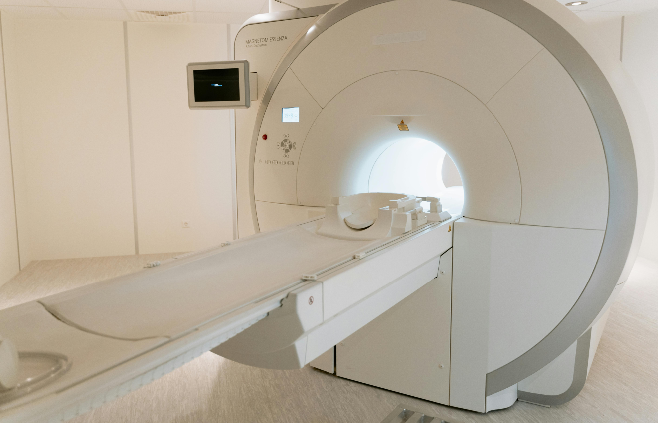
Multiparametric Contrast Brain MRIThe definitive gold standard for soft-tissue anatomical resolution; utilizes intravenous Gadolinium contrast with T1, T2, and FLAIR sequences.A specialized brain MRI delineates clear tumor margins, distinguishes active neoplastic tissue from surrounding vasogenic edema, and assesses ventricular compression.
Magnetic Resonance Spectroscopy (MRS)Non-invasive biochemical profiling measuring cellular metabolic markers within the tumor core.Detects elevated Choline-to-Creatine ratios (indicating rapid cellular turnover) and decreased N-acetylaspartate (NAA, reflecting neuronal loss).
Functional MRI (fMRI) & DTI TractographyPre-operative functional mapping; superimposes motor, sensory, and language pathways onto 3D structural imaging.Visualizes white matter tracts (corticospinal tract, arcuate fasciculus) to ensure maximal safe resection without damaging eloquent brain cortex.
Stereotactic Needle / Open BiopsyImage-guided tissue extraction from deep-seated or inoperable lesions using frameless neuronavigation.Provides definitive tissue specimens for light microscopy histopathology and molecular immunohistochemistry.

Molecular Biomarkers in Modern Neuro-Oncology

Under the World Health Organization (WHO) CNS5 Classification, brain tumors are categorized not only by their microscopic appearance but also by their molecular and genetic signatures:

  • Isocitrate Dehydrogenase (IDH1 / IDH2) Mutation Status: Distinguishes primary aggressive glioblastomas (IDH-wildtype) from lower-grade astrocytomas and oligodendrogliomas (IDH-mutant), which carry a significantly more favorable prognostic trajectory.
  • 1p/19q Co-Deletion: Hallmark genetic marker confirming an oligodendroglioma lineage; predicts high therapeutic sensitivity to alkylating chemotherapy regimens.
  • MGMT Promoter Methylation: Epigenetic silencing of the MGMT DNA-repair gene; predicts enhanced tumor response and improved survival with Temozolomide (TMZ) chemotherapy.
  • EGFR Amplification & TERT Promoter Mutations: Key molecular indicators of high-grade, aggressive tumor biology that guide the selection of targeted clinical protocols.

Why Advanced Imaging Precedes Every Surgical Plan

  • Differentiating Tumor from Edema: Standard scans often make peritumoral swelling look identical to tumor mass. T2-FLAIR MRI sequences isolate infiltrative malignant margins from reactive fluid buildup.
  • Vascular Mapping: Contrast magnetic resonance angiography (MRA) and venography (MRV) map vital surrounding blood vessels (such as the internal carotid artery or superior sagittal sinus) to prevent intra-operative vascular injury.
  • Assessing Surgical Resectability: High-resolution 3D reconstructions allow neurosurgeons to simulate surgical approaches, determine craniotomy entry points, and plan safe surgical corridors before entering the operating suite.

The Molecular Standard: In modern neuro-oncology, imaging identifies where the tumor is, but molecular genetics dictates how it behaves. Integrating high-resolution contrast imaging with genetic biomarkers ensures personalized, targeted treatment for every patient.

Modern Multimodal Management: Neurosurgery, Radiotherapy & Targeted Therapies

Managing intracranial neoplasms requires an aggressive, highly coordinated multidisciplinary strategy. As brain tumors infiltrate complex neural networks, effective treatment combines precision surgical cytoreduction, conformal radiation techniques, and molecularly targeted systemic therapies.

Choosing the right combination of brain tumor treatment options depends on tumor histology, WHO grade, molecular biomarkers (IDH, MGMT status), anatomical location, and the patient’s baseline performance status.

Surgical Oncology: The Cornerstone of Treatment

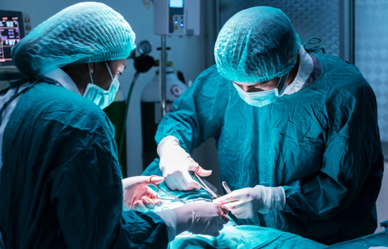
For the vast majority of accessible intracranial lesions, neurosurgery for tumors serves as the initial, definitive therapeutic intervention. The primary surgical goal is maximal safe resection excising the greatest possible volume of tumor while preserving intact neurological function.

Modern microsurgical suites employ advanced intraoperative technologies to maximize surgical accuracy:

  • Frameless Stereotactic Neuronavigation: Acts as a real-time intraoperative GPS, tracking surgical instruments in relation to 3D pre-operative MRI scans with sub-millimeter precision.
  • Intraoperative Neuromonitoring (IONM): Continuously records motor evoked potentials (MEP) and somatosensory evoked potentials (SSEP) to safeguard critical motor pathways throughout tumor dissection.
  • Awake Craniotomy & Cortical Mapping: For tumors located directly within or adjacent to eloquent language zones (Broca’s or Wernicke’s areas), patients are gently awakened intraoperatively to perform real-time speech and cognitive tasks, ensuring zero damage to speech centers during resection.

Fluorescence-Guided Surgery (5-ALA): Oral administration of 5-aminolevulinic acid causes malignant glioma cells to synthesize fluorescent protoporphyrin IX, illuminating infiltrative tumor margins in bright pink-red under specialized violet-blue operative microscope light.

Comprehensive Multimodal Treatment Modalities

Treatment ModalityClinical Mechanism & TechniquePrimary Indications & Clinical Value 
Maximal Safe Surgical ResectionMicrosurgical craniotomy aimed at Gross Total Resection (GTR) of tumor mass.Instantly relieves elevated intracranial pressure (ICP), eliminates mass effect, and provides extensive tissue for molecular staging.
Stereotactic Radiosurgery (SRS / CyberKnife)Delivers high-dose, sub-millimeter targeted radiation beams to well-defined lesions in 1 to 5 sessions.Primary choice for brain metastases, deep-seated recurrent tumors, vestibular schwannomas, and surgically inaccessible lesions.
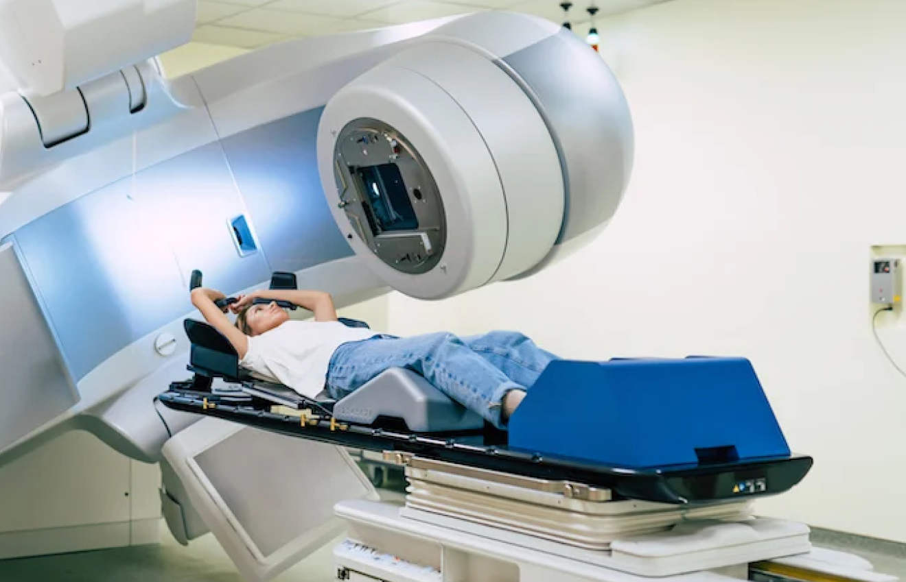
Intensity-Modulated Radiation Therapy (IMRT)Delivers fractionated, conformal photon beams shaped to the exact geometry of the tumor bed while sparing adjacent optic nerves and brainstem.Standard adjuvant local control for high-grade gliomas, astrocytomas, and atypical meningiomas following surgical resection.
Alkylating Chemotherapy (Temozolomide – TMZ)Oral lipophilic DNA-alkylating agent that efficiently crosses the Blood-Brain Barrier (BBB).The cornerstone of the standard Stupp Protocol for Glioblastoma (concomitant with radiotherapy, followed by adjuvant cycles).
Anti-Angiogenic & Targeted TherapyMonoclonal antibodies (e.g., Bevacizumab/VEGF inhibitors) and mutant-enzyme blockers (IDH inhibitors).Rapidly resolves refractory vasogenic edema, controls tumor vascularity, and targets specific molecular driver mutations.
Supportive Neuro-Intensive CareHigh-dose corticosteroids (Dexamethasone), osmotic agents (Mannitol), and modern antiepileptics (Levetiracetam).Rapidly reduces peritumoral swelling, controls acute intracranial hypertension, and prevents debilitating seizure activity.

The Power of the Multidisciplinary Tumor Board

No single specialist manages brain tumors in isolation. Modern neuro-oncology relies on a collaborative Multidisciplinary Tumor Board (MDT):

  • Surgical Oncologists & Neurosurgeons: Determine resectability, execute microsurgical decompression, and preserve functional neurological architecture.
  • Medical Oncologists: Design systemic chemotherapy, targeted therapy, and clinical trial regimens based on somatic mutation profiling.
  • Radiation Oncologists: Plan computer-optimized radiation target volumes to prevent recurrence in the surrounding infiltrative margin.
  • Neuro-Pathologists: Perform high-resolution immunohistochemistry and molecular cytogenetics to confirm WHO grading.
  • Neuro-Rehabilitation Specialists: Initiate early post-operative physiotherapy, speech therapy, and occupational therapy to restore physical independence.

Total tumor removal must never come at the cost of permanent neurological deficit. Modern neuro-oncology focuses on achieving the optimal balance between maximizing tumor resection volume and preserving the patient’s long-term functional independence and quality of life.

Comprehensive Neuro-Oncology Care at Dasappa Cancer Hospital

Managing complex intracranial lesions demands surgical precision, rapid diagnostic access, and dedicated oncological infrastructure.

At Dasappa Cancer Hospital (Dasappa Memorial Institute of Oncology), our specialized oncology and neuro-care teams provide end-to-end diagnostic evaluations, surgical coordination, and customized adjuvant systemic protocols within a focused, compassionate hospital environment.

Under the clinical leadership of Dr. Pandu Dasappa (M.Ch Surgical Oncology, GCRI Ahmedabad; FIAGES, FMAS; with over 27 years of surgical and clinical oncology experience), our multidisciplinary teams integrate surgical onco-resection principles, advanced histopathology, and evidence-based medical oncology to deliver optimal patient outcomes.

Core Hospital Strengths & Clinical Infrastructure

  • Dedicated 75-Bed Super-Speciality Oncology Facility: Purpose-built inpatient wards, surgical step-down units, and high-dependency intensive care units (ICU) equipped for continuous neurological monitoring, intracranial pressure management, and acute post-operative stabilization.
  • Multidisciplinary Tumor Board (MDT) Governance: Every brain tumor presentation is evaluated by a collaborative panel of surgical, medical, radiation, and diagnostic specialists to ensure staging and therapeutic protocols align with NCCN and ICMR clinical guidelines.
  • Specialized Day-Care Infusion Suites: Modern, comfortable day-care chemotherapy suites staffed by certified oncology nursing personnel for delivering targeted biological therapies, anti-angiogenic infusions (such as Bevacizumab), and standard adjuvant protocols.
  • Rapid Diagnostic & Pathology Coordination: Expedited processing of high-resolution contrast neuro-imaging, stereotactic biopsies, and advanced molecular markers (IDH1/2, 1p/19q, MGMT methylation) to eliminate critical treatment delays.
  • Integrated Neuro-Rehabilitation & Supportive Care: Early mobilization protocols combining specialized physical therapy, occupational therapy, speech and swallowing rehabilitation (dysphagia management), clinical oncology nutrition, and psychological counseling for patients and their families.

The Dasappa Patient-First Care Pathway

  • Prompt Clinical Triage: Rapid appointments and imaging reviews for patients presenting with acute red-flag neurological signs or unprovoked adult seizures.
  • Transparent Treatment Planning: Clear, compassionate discussions detailing expected surgical goals, functional risk mitigation, and step-by-step adjuvant therapy roadmaps with no hidden procedural costs.
  • Long-Term Surveillance: Structured follow-up imaging protocols and cognitive monitoring to detect potential disease recurrence early and maintain functional independence.

Combining surgical experience, multi-specialty collaboration, and dedicated inpatient oncology support ensures that every patient receives focused, scientifically grounded care designed to preserve neurological function and optimize long-term survival.

Restoring Hope Through Early Intervention

Intracranial space-occupying lesions represent complex clinical challenges, but early symptom recognition fundamentally alters the treatment trajectory. Identifying warning signs such as unprovoked adult seizures, progressive early-morning headaches, sudden speech hesitations, or focal motor weakness enables rapid imaging and intervention before elevated intracranial pressure causes irreversible neurological damage.

Early neurosurgical decompression, combined with precision molecular profiling and conformal adjuvant therapies, preserves functional independence, protects cognitive baseline, and significantly improves long-term survival outcomes.

Consultation & Clinical Assessment Details

If you, a family member, or a patient is experiencing persistent neurological changes, uncharacteristic headaches, or abnormal brain imaging findings, reach out to our specialized oncology team in Bengaluru for an immediate evaluation:

  • Hospital Location: Dasappa Memorial Institute of Oncology (Dasappa Cancer Hospital), No. 132, Gandhi Bazaar Main Road, Basavanagudi, Bengaluru, Karnataka 560004
  • Consultation Helplines: +91 80 2667 9294 / +91 98450 12345
  • Website: dasappacancerhospital.com

Specialized Oncology Services: Surgical Oncology, Medical Oncology, Chemotherapy & Targeted Infusion Suites, Histopathology & Molecular Diagnostics, Second Opinion Consultations.

About The Author

Share With Your Network

Recent Posts

Book An Appointment