




Top Benefits of IMRT Radiotherapy for Cancer Patients

Table of Contents

Receiving a cancer diagnosis can be an overwhelming moment filled with intense uncertainty regarding the medical journey ahead.

When exploring options for localized advanced care, understanding the therapeutic tools available can fundamentally change your outlook on recovery. Utilizing IMRT in radiotherapy represents a clinical leap forward, offering a highly precise approach that treats target tissue while saving neighboring healthy anatomy.

Getting an evaluation at the Best cancer hospital in Bangalore provides individuals with direct access to this advanced, customized technology, turning a complicated medical crossroad into a clear pathway toward long-term survival.

Historically, a major challenge in radiation oncology was the balance between delivering a dose strong enough to eradicate malignant cells and avoiding toxic exposure to adjacent vital organs. 

Standard legacy approaches often applied static, uniform blocks of energy, which could inadvertently harm healthy structural layers. Modern therapeutic design addresses this tension by dynamically altering the shape and intensity of the treatment field.

We at Dasappa Cancer Hospital frame advanced oncological screening and treatment around patient longevity and post-therapy quality of life, empowering families with transparent education so they can confidently master their recovery milestones.

Medical Disclaimer

The clinical insights, technical breakdowns, and treatment outcomes presented in this guide are intended strictly for educational and informational purposes. This content does not constitute professional medical advice, a formal oncological diagnosis, or a definitive clinical treatment plan. Tumor shapes, internal anatomy, and radiation tolerance thresholds are highly unique biological factors that vary significantly based on genetics, medical history, and age. Always consult a qualified radiation oncologist or a board-certified medical specialist to evaluate your specific parameters before beginning any advanced therapy protocol. Never delay seeking professional medical care based on the material read in this article.

The Technological Shift: How Intensity Modulation Redefines Targeting?

To understand why modern radiation oncology has advanced so significantly, it helps to examine how the delivery of radiation beams has changed over time. For many years, conventional radiation therapy relied on three-dimensional conformal systems (3D-CRT).

While 3D-CRT allowed oncologists to shape radiation beams to match the physical outline of a tumor, the strength of the beam remained completely uniform across the entire treatment field. This meant that healthy tissues lying directly in front of or behind the malignancy received the same concentration of radiation as the core of the tumor itself.

This is where intensity-modulated radiation therapy marks a critical structural shift in oncological treatment. Instead of treating the tumor with a single, uniform wall of energy, this advanced technique breaks a single radiation beam down into thousands of microscopic, individual “beamlets.”

Adjusting the power of each beamlet independently, clinical teams can sculpt the radiation to curve precisely around the complex, irregular shapes of a tumor.

The Mechanics of Real-Time Shaping

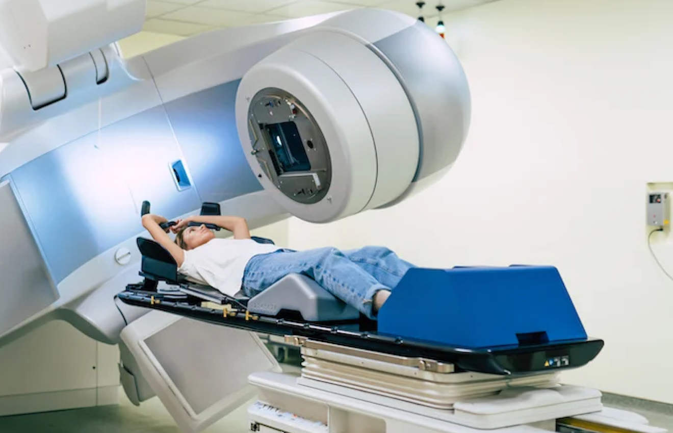
This highly precise tailoring is made possible by a specialized mechanical component within the linear accelerator known as a Multi-Leaf Collimator (MLC).

  • Dynamic Micro-Leaves: The MLC consists of up to 120 computer-controlled tungsten leaves arranged in matching pairs. As the machine rotates around the patient, these metal leaves slide rapidly back and forth in real-time, constantly altering the shape of the beam opening.
  • Varying Power Levels: By selectively blocking parts of the beam while allowing others to pass through, the leaves dynamically vary the intensity of the radiation across the treatment field.

Areas of the tumor requiring aggressive treatment receive a concentrated dose, while sections bordering critical healthy structures receive a heavily reduced exposure.

The Power of Inverse Planning Software

The true intelligence behind this approach lies in a digital configuration process called inverse planning. In conventional therapies, a dosimetrist manually estimates the beam angles and checks if the healthy tissue is safe. In contrast, inverse planning flips the workflow completely:

Clinical Workflow: The oncology team first defines the precise, lethal radiation dose needed to destroy the tumor, alongside the absolute maximum safety thresholds allowed for surrounding vital structures. Specialized computer software then runs complex optimization algorithms, performing thousands of calculations to determine the exact angles, beam intensities, and MLC movements required to achieve that specific clinical outcome safely.

This transition from manual estimation to digital optimization ensures that the treatment is entirely custom-tailored to your unique internal anatomy. By transforming a uniform block of radiation into a highly flexible, variable energy field, this system gives clinical teams the power to target difficult malignancies with remarkable accuracy while protecting the structural health of your body.

Clinical Superiority of Minimizing Toxicity and Maximizing Dose Conformity

The true breakthrough of this targeted methodology is its ability to handle two opposing clinical goals simultaneously: dose escalation and toxicity mitigation. In traditional oncological treatments, increasing the radiation dose to maximize the destruction of tumor cells inherently meant increasing the damage to neighboring healthy structures.

Utilizing advanced dynamic planning, clinical teams can safely elevate the radiation volume inside the tumor boundary while drastically lowering the exposure to nearby healthy organs. This architectural shift provides two major clinical advantages that fundamentally redefine the patient treatment path.

1. Dose Escalation Within Complex Volumes

Higher Radiation Precision

Tumors rarely grow in neat, symmetrical spheres. They often wrap around critical internal structures like the spinal cord, brainstem, or major blood vessels.

When delivering a modern IMRT radiotherapy protocol, clinicians can precisely shape the radiation field to deliver an intense, destructive dose directly to the malignant core while creating sharp, steep drops in energy lines right at the border of sensitive tissues.

For example, in pelvic treatments, the system allows the delivery of higher doses, often exceeding 76 Gy to 78 Gy directly to the target area while keeping the surrounding rectal and bladder tissue well within safe exposure limits.

2. Radical Reduction in Side Effects

Fewer Side Effects

By systematically avoiding healthy organs at risk, this targeted methodology mitigates many of the painful, life-altering toxicities traditionally associated with high-dose radiation.

Data from landmark randomized clinical trials highlight the real-world impact of this targeting precision:

1. Head and Neck Preservation: In head and neck malignancies, conventional uniform radiation frequently damages the parotid salivary glands, causing permanent xerostomia (severe chronic dry mouth).

Long-term clinical data show that while 83% of patients undergoing conventional static treatments suffer from severe long-term dry mouth two years post-therapy, that number drops to just 29% for patients who receive a parotid-sparing modulated protocol.

2. Gastrointestinal Protection: For patients dealing with localized pelvic malignancies, mapping the exact structural boundaries reduces overall moderate-to-severe acute gastrointestinal complications from 49% to 29%.

Furthermore, the 5-year risk of developing late gastrointestinal complications drops from 20% to just 8%.

The Quality-of-Life Factor: Minimizing this internal structural damage means patients experience far less pain, fewer nutritional challenges, and a significantly lower risk of tissue scarring during their treatment journey.

Choosing a highly optimized IMRT treatment ensures that the aggressive destruction of cancer cells does not come at the cost of your long-term, post-treatment physical well-being.

The Metrics of Efficacy and Evaluating Clinical Outcomes

When oncology teams analyze the clinical impact of advanced treatment tracking, success is measured using two distinct variables: localized tumor control (the complete eradication of targeted cancer cells) and organ preservation (the long-term structural health of the surrounding functional tissue).

As legacy systems often restricted the total radiation dose due to the risk of surrounding tissue damage, tumor clearance rates could be limited in complex or deep-seated cases. By shifting to a variable-intensity delivery model, clinical data demonstrates an impressive upward shift in both patient survival metrics and lifestyle preservation.

Evaluating long-term oncology registry data across thousands of international cases reveals that this targeting precision translates directly into superior therapeutic outcomes.

Comparative Analysis of Clinical Efficacy

Clinical Effectiveness

The table below breaks down the differences in treatment performance between conventional uniform radiation fields (3D-CRT) and advanced modulated delivery across primary oncological sites:

Malignancy SiteClinical Evaluation ParameterConventional 3D-CRT OutcomesAdvanced Modulated Delivery Outcomes 
Head & Neck Cancers3-Year Localized Tumor Control Rate78% localized clearance86% localized clearance
Long-Term Salivary Function PreservationLess than 20% of patients retain normal salivaGreater than 65% of patients retain normal saliva
Prostate Cancer5-Year Biochemical Disease-Free Survival82% success rate in early-to-mid stages92% success rate due to safe dose escalation
Long-Term Late Rectal Toxicity Rate15% to 20% experience chronic inflammationLess than 5% experience chronic inflammation
Gynecological MalignanciesAcute Hematological (Bone Marrow) Toxicity55% require treatment interruption30% require treatment interruption
Pelvic Insufficiency Fracture Risk12% experience long-term bone weaknessLess than 4% experience long-term bone weakness
Thoracic (Lung) TumorsIncidence of Severe Radiation Pneumonitis18% suffer acute lung lining inflammation8% suffer acute lung lining inflammation

Deconstructing the Clinical Performance Data

A deep review of modern clinical oncology records reveals that the IMRT success rate is highly evident in specific, highly sensitive anatomical zones:

1. Dose Escalation Efficacy: In prostate cancer treatment, conventional radiotherapy methods often capped the total target dose at 70 Gy to prevent severe, permanent damage to the adjacent rectal wall. With Intensity-Modulated Radiation Therapy (IMRT), clinicians can safely escalate the dose to 78 Gy or even 80 Gy, delivering more effective treatment to the prostate while minimizing radiation exposure to nearby healthy tissues. 

This significant increase in energy delivery provides a 10% gain in long-term biochemical control, preventing cancer recurrence without increasing patient complications.

2. Protection of Critical Biological Structures: In brain and skull-base treatments, tumors often sit directly against the optic nerves or the brainstem.

The sharp dose fall-off achieved with modern computer-driven leaves ensures that these vital neurological pathways receive an exposure well below their structural tolerance limits, eliminating the risk of blindness or central nervous system damage.

3. Maintenance of Active Treatment Timelines: Traditional wide-field radiation frequently hits the pelvic bone marrow, leading to sharp drops in white blood cell counts (leukopenia). When a patient’s blood counts drop too low, active cancer therapy must be temporarily paused to let the body recover, allowing the tumor to regenerate. 

Modulated pathways carefully bypass the active bone marrow structures within the pelvic girdle.

This structural protection keeps blood counts stable, ensuring patients complete their full treatment cycles on schedule without unnecessary, dangerous delays.

Why Choose Dasappa Cancer? Precision Oncological Care at Dasappa Cancer Hospital

Choosing where to undergo specialized oncology treatment is a pivotal decision that directly shapes your recovery timeline and therapeutic experience. For individuals seeking advanced radiation therapy in Bangalore, Dasappa Cancer Hospital provides an integrated, world-class clinical ecosystem designed to deliver maximum targeting accuracy and compassionate care under one roof.

Our oncology department is engineered around these core operational capabilities:

  • Advanced Linear Accelerator Suites: Equipped with cutting-edge tracking infrastructure that dynamically shapes energy beams to conform exactly to irregular tumor boundaries.
  • Expert Dosimetry and Planning Panel: A dedicated team of clinical medical physicists who utilize state-of-the-art digital software to run complex inverse planning algorithms for every patient.
  • Proactive Toxicity Management: Rigorous, daily monitoring protocols specifically designed to safeguard adjacent healthy organs and maximize your comfort during active treatment.

A collaborative clinical environment connecting radiation specialists, surgical oncologists, and nutritionists to manage your entire wellness continuum.

Conclusion

Advanced target shaping represents a fundamental shift in modern oncology, moving away from rigid, wide-field radiation blocks toward highly customized, variable-intensity care. By utilizing these computer-driven protocols, medical teams can aggressively target deep-seated malignancies while protecting the structural integrity of the surrounding healthy tissue.

Ultimately, this technical evolution means that fighting cancer no longer requires sacrificing your long-term physical well-being. Partnering with a dedicated clinical team ensures that your treatment path is optimized for both maximum cell eradication and the preservation of an active, fulfilling life after recovery.

About The Author

Share With Your Network

Recent Posts

Book An Appointment