

Recognizing the subtle, early signs of blood cancer is critical for timely diagnostic triage and achieving favorable long-term remission.
Unlike solid-organ tumors that typically form localized, palpable masses, hematologic malignancies originate deep within the bone marrow and lymphatic system, circulating quietly throughout the body.
When persistent fatigue, unexplained bruising, or recurrent infections arise, seeking comprehensive clinical evaluation for Blood Cancer Treatment in Bangalore ensures access to advanced oncological diagnostics, multidisciplinary tumor boards, and specialized therapies designed to halt disease progression before acute complications occur.
Blood cancers encompassing leukemias, lymphomas, and multiple myeloma arise from disruptions in hematopoiesis, the biological process responsible for generating mature blood cells. Within the spongy interior of bone marrow, pluripotent stem cells differentiate into red blood cells (erythrocytes), white blood cells (leukocytes), and platelets (thrombocytes).
As these early manifestations closely mimic everyday viral infections, chronic stress, or benign nutritional deficiencies (such as iron or Vitamin B12 deficiency), diagnosis is often delayed until routine blood work reveals significant abnormalities.
What You Will Discover in This Guide
- Triaging the 3 Major Classes: Differentiating the clinical patterns of Leukemias, Lymphomas, and Multiple Myeloma.
- The Precision Diagnostic Pathway: Understanding the diagnostic journey from a Complete Blood Count (CBC) and peripheral blood smear to bone marrow biopsy and flow cytometry.
- Benign Blood Disorders vs. Malignancy: Learning how to distinguish reactive conditions from true hematologic neoplasms.
- Modern Oncological Therapies: How early detection enables targeted therapies, immunotherapy, and multi-agent regimens to achieve durable disease control.
Accurate hematologic evaluation demands sharp clinical acumen, swift onco-pathology turnaround, and an integrated multidisciplinary approach. Led by Dr. Pandu Dasappa (M.Ch Surgical Oncology, GCRI Ahmedabad; 27+ years of surgical and clinical oncology experience), Dasappa Cancer Hospital provides evidence-based, compassionate care, ensuring patients receive precise diagnostic clarity and tailored systemic therapy protocols.
Medical Disclaimer
The hematologic descriptions, diagnostic pathways, and treatment overviews presented in this article are intended strictly for educational and public health awareness purposes. This content does not constitute formal medical advice, clinical diagnosis, or a personalized oncology management plan. Any blood count abnormalities, marrow evaluations, or therapeutic decisions must be evaluated by a qualified medical oncologist, hematologist, surgical oncologist, or licensed physician.
Triaging the 3 Major Classes: Leukemia, Lymphoma & Multiple Myeloma
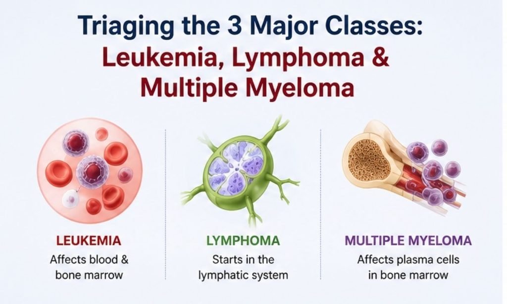
Hematologic malignancies are categorized into three broad pathological classifications based on the cell lineage and anatomical site of origin. As each subtype affects a different component of the blood-forming or immune systems, the clinical presentation and specific blood cancer symptoms vary substantially between patients.
Hematologic Malignancy Triage Matrix
| Disease Category | Primary Cellular Origin | Hallmark Early Clinical Manifestations | Key Differentiating Pathophysiology |
|---|---|---|---|
| Leukemia(AML, ALL, CML, CLL) | Bone marrow hematopoietic stem cells (Myeloid or Lymphoid blasts). | • Extreme, unrelenting fatigue and pallor • Spontaneous bruising, petechiae, and gum bleeding • Recurrent fevers, mouth sores, and persistent infections • Deep, aching bone and joint pain | Rapid clonal expansion of immature blast cells within marrow cavities suppresses normal red cell, white cell, and platelet production. |
| Lymphoma(Hodgkin & Non-Hodgkin) | Mature lymphocytes in peripheral lymph nodes and lymphatic organs. | • Painless, rubbery swelling in neck, armpits, or groin • Drenching night sweats requiring bedding changes • Unexplained weight loss (> 10% in 6 months) • Persistent generalized skin itching (pruritus) | Malignant lymphocytes accumulate within lymphoid tissue, forming solid nodal aggregates and releasing pyrogenic cytokines. |
| Multiple Myeloma | Antibody-producing plasma cells in the bone marrow. | • Persistent, unprovoked lower back and rib pain • Chronic fatigue and microcytic/normocytic anemia • Unexplained kidney dysfunction or foamy urine • Recurrent respiratory or urinary tract infections | Clonal plasma cells overproduce non-functional monoclonal immunoglobulin (M-protein) and stimulate osteoclastic bone destruction. |
1. Leukemias: Acute vs. Chronic Presentations
Leukemias represent malignancies of the blood-forming tissues and are broadly divided by their rate of progression:
- Acute Leukemias (AML / ALL): Characterized by an abrupt, rapid proliferation of non-functional immature blast cells. Symptoms develop rapidly over days to weeks, typically presenting with severe fatigue, unexplained subcutaneous bleeding (petechiae), and stubborn infections that fail to respond to standard antibiotics.
- Chronic Leukemias (CML / CLL): Progress insidiously over months or years. Mature but dysfunctional leukocytes accumulate slowly; many patients remain asymptomatic initially, with the condition discovered incidentally via elevated white blood cell counts on routine blood panels or during an evaluation for early satiety caused by an enlarged spleen.
2. Lymphomas: Nodal Enlargement & Constitutional B-Symptoms
Lymphomas develop when lymphocytes proliferate abnormally within the lymph nodes, spleen, thymus, or gastrointestinal tract:
- Painless Lymphadenopathy: The classic presentation is one or more firm, movable, painless enlarged lymph nodes in the cervical (neck), axillary (armpit), or supraclavicular regions that persist for more than 4 to 6 weeks without an obvious underlying localized infection.
- Constitutional B-Symptoms: Approximately 30% of patients experience systemic cytokine-driven signs specifically unexplained fevers exceeding 38°C (100.4°F), drenching night sweats, and rapid unintentional weight loss which directly indicate systemic disease activity.
3. Multiple Myeloma & The CRAB Clinical Criteria
Multiple Myeloma is a disease of mature plasma cells that primarily affects older adults. Clinicians identify early organ compromise using the established CRAB diagnostic criteria:
- C – Hypercalcemia: Bone destruction releases excess calcium into the blood, causing mental confusion, extreme thirst, constipation, and muscle weakness.
- R – Renal Impairment: Excess circulating light-chain proteins (Bence Jones proteins) precipitate in renal tubules, leading to progressive kidney strain and elevated serum creatinine.
- A – Anemia: Plasma cell crowding within the marrow leads to decreased erythropoiesis and chronic fatigue.
- B – Bone Lytic Lesions: Accelerated osteoclast activity causes targeted osteolytic punched-out bone lesions, severe skeletal pain (especially in the spine and ribs), and pathological fractures resulting from minimal trauma.
Distinguishing Malignancies from Benign Blood Disorders
Non-malignant, reactive blood disorders are far more common than hematologic cancers and can present with overlapping signs:
- Nutritional Anemia vs. Marrow Failure: Iron deficiency or Vitamin B12 deficiency causes fatigue and pallor, but responds rapidly to nutritional repletion and typically spares white blood cell and platelet counts.
- Reactive Lymphadenitis vs. Lymphoma: Viral or bacterial throat infections frequently cause tender, painful, soft lymph node swelling that resolves completely within 2 to 3 weeks once the primary infection clears.
- Benign Thrombocytopenia (ITP) vs. Leukemia: Immune thrombocytopenic purpura causes isolated low platelet counts without the blast cells, profound leukopenia, or severe anemia seen in malignant bone marrow disorders.
Benign symptoms generally fluctuate or resolve as acute infections subside. Any progressive, painless lymph node enlargement, persistent unexplained fever, or unprovoked bruising lasting beyond 3 to 4 weeks warrants targeted hematologic investigation.
The Precision Diagnostic Roadmap: From CBC to Bone Marrow Biopsy
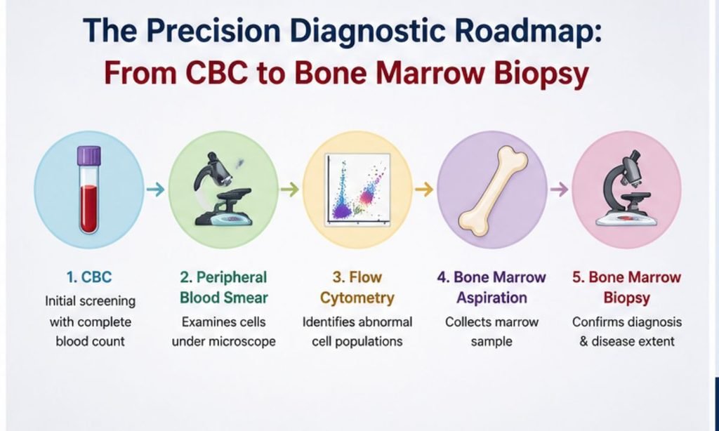
Establishing an accurate blood cancer diagnosis requires a step-by-step diagnostic cascade that transitions from routine venous blood tests to specialized cellular and molecular genetic analyses.
As hematologic malignancies display substantial biological variation, identifying the exact clonal lineage, mutation profile, and cellular maturity dictates the precise choice of induction chemotherapy, targeted small-molecule inhibitors, or immunotherapy.
The 4-Tier Diagnostic Cascade
| The Hematologic Oncology Diagnostic Cascade |
|---|
| 1. Complete Blood Count (CBC) & Automated Differential |
| 2. Manual Peripheral Blood Smear Examination |
| 3. Bone Marrow Aspiration & Trephine Core Biopsy |
| 4. Flow Cytometry (Immunophenotyping) & Molecular Cytogenetics (FISH/NGS) |
1. Initial Screening: CBC & Peripheral Blood Smear
The diagnostic evaluation begins with basic peripheral blood analysis:
Complete Blood Count (CBC): Identifies quantitative abnormalities across blood lines, including profound anemia, severe thrombocytopenia (< 50,000/µL), unexplained neutropenia, or marked leukocytosis (> 50,000 to 100,000/µL).
Manual Peripheral Smear: A hematopathologist examines cellular morphology under a light microscope to evaluate red cell shape, detect abnormal platelet clumps, and look for circulating immature blast cells (which are normally confined entirely to the bone marrow) or distinctive morphological features such as Auer rods in myeloblasts.
2. Definitive Tissue Evaluation: Bone Marrow Aspiration & Biopsy
When peripheral blood findings or persistent unexplained cytopenias suggest marrow failure, a bone marrow examination serves as the gold standard confirmatory test:
Bone Marrow Aspiration: A fine needle draws liquid marrow from the posterior superior iliac crest (pelvic bone) to assess cellular morphology, differential blast count, and maturation stages under high magnification.
Trephine Core Biopsy: A small, solid core of spongy bone is extracted to evaluate overall marrow cellularity, architectural disruption, fibrosis, and plasma cell infiltration patterns.
3. Advanced Molecular & Genetic Profiling
Modern hemato-oncology relies on high-resolution genetic and immunological testing to classify disease subtypes and stratify prognostic risk:
| Diagnostic Modality | Analytical Purpose | Key Clinical Output |
|---|---|---|
| Flow Cytometry (Immunophenotyping) | Analyzes cell-surface and cytoplasmic Cluster of Differentiation (CD) protein markers using laser fluorometry. | Distinguishes B-cell vs. T-cell lymphoid lineages, confirms myeloid markers (CD13, CD33, MPO), and detects minimal residual disease (MRD). |
| Cytogenetics (Karyotyping) | Examines structural chromosomal rearrangements in dividing malignant cells. | Identifies characteristic chromosomal translocations, such as Philadelphia chromosome t(9;22) in CML/ALL or t(15;17) in Acute Promyelocytic Leukemia (APL). |
| Fluorescence In Situ Hybridization (FISH) | Uses fluorescent DNA probes to detect specific micro-deletions or gene fusions. | Detects high-risk genetic lesions, including del(17p), TP53 mutations, or MYD88/IGH rearrangements in multiple myeloma and lymphoma. |
| Next-Generation Sequencing (NGS) | Comprehensive multi-gene panel sequencing for targetable somatic mutations. | Identifies actionable driver mutations such as FLT3, NPM1, IDH1/2, and BRAF to guide precision targeted therapy. |
4. Staging Imaging: PET-CT & Skeletal Assessments
To determine the anatomical extent of disease outside the bone marrow cavity:
- Whole-Body 18F-FDG PET-CT: Serves as the primary staging and restaging modality in Hodgkin and Non-Hodgkin Lymphomas to map hypermetabolic lymph node stations and extra-nodal organ involvement.
- Low-Dose Whole-Body CT / Skeletal Survey: Identifies cortical bone thinning, osteolytic lesions, and pathological fracture risks in patients diagnosed with Multiple Myeloma.
The Pathological Benchmark: A complete hematologic workup does not simply identify the presence of blood cancer; it maps the exact chromosomal and molecular profile of the disease, ensuring oncologists can select the most effective targeted and systemic therapies.
Why Blood Cancer Lacks Routine Tests
For solid-organ malignancies, established population-level screening programs such as digital mammography for breast cancer, Pap smears and HPV DNA testing for cervical cancer, and optical colonoscopy for colorectal polyps allow clinicians to detect pre-malignant lesions years before invasive transformation.
However, in hematologic oncology, routine asymptomatic cancer screening protocols do not exist in the traditional sense.
Understanding why blood cancers lack standardized screening tests highlights the critical importance of clinical vigilance, symptom awareness, and prompt diagnostic investigation when unexplained blood count abnormalities arise.
Why Standard Screening Modalities Do Not Apply to Liquid Tumors
The biological differences between solid-organ neoplasms and hematologic malignancies explain why population-wide screening remains a challenge:
- Absence of a Localized Pre-Invasive Phase: Solid tumors typically progress through identifiable pre-malignant stages (such as ductal carcinoma in situ or adenomatous colonic polyps) confined to a specific tissue layer. In contrast, hematologic malignancies originate in the bone marrow cavity and can enter systemic circulation almost immediately upon emergence.
- Rapid Cellular Doubling Times: Acute leukemias in particular feature rapid blast cell proliferation. A patient with a completely normal Complete Blood Count (CBC) during an annual health checkup can develop acute leukemia symptoms weeks later, rendering infrequent interval screening ineffective.
As blood and lymphatic channels span every organ, there is no single localized anatomical target (such as the cervix or colon) that can be visualized or biopsied non-invasively during a routine physical exam.
Solid Tumor Screening vs. Hematologic Diagnostic Vigilance
| Screening Dimension | Solid-Organ Malignancies | Hematologic Malignancies (Blood Cancers) |
|---|---|---|
| Primary Screening Method | Mammography, Pap smear, Colonoscopy, Low-Dose CT (LDCT). | No routine population test; relies on clinical vigilance and opportunistic blood work. |
| Detectable Pre-Malignant Stage | In-situ lesions, dysplastic mucosal polyps, atypical hyperplasia. | Rare (e.g., MGUS in Myeloma, Clonal Hematopoiesis of Indeterminate Potential – CHIP). |
| Screening Trigger | Age-based or population-risk milestones (e.g., annual screening after age 40/45). | Symptom-triggered investigation, persistent cytopenias, or abnormal routine CBC. |
| Definitive Confirmation | Core needle biopsy or surgical excision of the primary solid mass. | Bone marrow aspiration/biopsy, peripheral blood smear, and flow cytometry. |
The Role of Opportunistic Screening & Routine Blood Audits
While population-wide mass screening is not standard, routine annual blood tests frequently serve as an opportunistic diagnostic safety net:
- The Routine CBC as a First Warning: Unexplained, persistent mild anemia, low platelet counts (thrombocytopenia), or elevated white blood cell counts with atypical differentials during annual checkups frequently provide the earliest clue of an underlying bone marrow disorder.
- Serum Protein Electrophoresis (SPEP): In adults over 50 presenting with persistent back pain, fatigue, or unexplained renal dysfunction, evaluating for a monoclonal M-spike can catch asymptomatic plasma cell dyscrasias such as Monoclonal Gammopathy of Undetermined Significance (MGUS) or Smoldering Multiple Myeloma prior to osteolytic bone destruction.
Any firm, non-tender, rubbery lymph node that remains enlarged for more than 4 to 6 weeks in the absence of an active infection warrants ultrasound mapping and core-needle or excisional biopsy.
When to Escalate to an Oncology Specialist
The key to early intervention lies in recognizing when common symptoms exceed expected recovery timelines:
- The 3-to-4-Week Persistence Rule: Benign, reactive conditions (such as viral pharyngitis, post-infectious fatigue, or minor iron deficiency) typically resolve or improve within 2 to 4 weeks with standard supportive care. Persistent, worsening symptoms warrant hematologic escalation.
- Multi-Lineage Cytopenias: If a blood test demonstrates abnormalities across two or more cell lines (e.g., anemia paired with low platelets or unexplained leukocytosis), an immediate peripheral blood smear and hematology/oncology consultation are strongly recommended.
- B-Symptoms with Normal Physical Exams: Unexplained fevers, drenching night sweats, and significant involuntary weight loss without a clear infectious source require specialized systemic oncological workup.
The Clinical Vigilance Rule: As blood cancers cannot be caught through routine solid-tumor imaging, prompt attention to abnormal blood counts and persistent systemic symptoms remains the single most effective tool for timely, life-saving diagnosis.
Modern Hemato-Oncology Therapies & The Power of Early Intervention

Over the past decade, the landscape of hematologic oncology has transitioned from non-selective cytotoxic regimens to precision-targeted molecular therapies, bioengineered antibodies, and cellular immunotherapies.
Today, the clinical success of these advanced treatments depends heavily on early cancer detection and immediate therapeutic initiation before malignant blast expansion triggers life-threatening systemic emergencies.
When blood cancers are caught early, patients present with lower baseline tumor burden, intact renal and hepatic function, and preserved immune reserves substantially reduce treatment-related toxicity and improve overall survival.
Preventing Life-Threatening Acute Complications
Delayed presentation of hematologic malignancies can lead to rapid medical crises that require immediate emergency stabilization before definitive anti-cancer therapy can even begin:
- Disseminated Intravascular Coagulation (DIC): Particularly in Acute Promyelocytic Leukemia (APL), malignant promyelocytes release procoagulant proteins that cause catastrophic microvascular clotting and life-threatening systemic hemorrhages. Early diagnosis allows prompt administration of All-Trans Retinoic Acid (ATRA) and Arsenic Trioxide (ATO), curing over 90% of cases without standard cytotoxic chemotherapy.
- Hyperleukocytosis & Leukostasis: Extreme white blood cell counts (> 100,000/µL) sludge small capillaries in the brain and lungs, risking intracranial hemorrhage and acute respiratory distress syndrome (ARDS).
- Tumor Lysis Syndrome (TLS): Rapid spontaneous lysis of massive tumor cells releases toxic levels of potassium, phosphorus, and uric acid into the bloodstream, triggering acute renal failure and fatal cardiac arrhythmias.
Prolonged bone marrow suppression leaves patients vulnerable to opportunistic fungal, viral, and bacterial pathogens.
The Modern Therapeutic Spectrum
Modern hematologic oncology relies on risk-adapted, multimodality protocols tailored to exact cytogenetic and molecular markers:
| Therapeutic Class | Mechanism of Action | Representative Clinical Indications | Key Therapeutic Advantage |
|---|---|---|---|
| Targeted Small-Molecule Inhibitors | Selectively blocks aberrant intracellular enzymes and signaling proteins (TKIs, BTK inhibitors, BCL-2 inhibitors). | • CML: Imatinib, Dasatinib • CLL/Mantle Cell: Ibrutinib, Acalabrutinib • AML: Venetoclax, FLT3 inhibitors | High disease control rates with oral administration; avoids severe marrow toxicity of traditional chemo. |
| Monoclonal Antibodies & BiTEs | Engineered antibodies that tag tumor-specific antigens (CD20, CD30, CD19) for immune destruction. | • B-Cell Lymphomas: Rituximab • Hodgkin Lymphoma: Brentuximab • B-ALL: Blinatumomab (Bi-specific T-cell engager) | Recruits the patient’s own cytotoxic T-cells directly to malignant lymphoid cells. |
| Multi-Agent Induction Chemotherapy | Cytotoxic combinations that rapidly clear blast populations to restore normal marrow hematopoiesis. | • AML (7+3 Cytarabine / Daunorubicin) • ALL (Multi-phase induction) • Aggressive Lymphomas (CHOP / EPOCH) | Induces deep complete morphological remission (CR) as a bridge to consolidation or transplantation. |
| Cellular Immunotherapy (CAR-T Cell Therapy) | Genetically modifies autologous T-cells with chimeric antigen receptors targeting CD19/CD22/BCMA. | • Relapsed/Refractory B-ALL • Diffuse Large B-Cell Lymphoma (DLBCL) • Relapsed Multiple Myeloma | Delivers durable complete remissions in heavily pre-treated, refractory hematologic disease. |
| Stem Cell Transplantation (BMT) | Replaces diseased marrow with healthy hematopoietic stem cells (Autologous or Allogeneic). | • High-risk AML / ALL in first remission • Multiple Myeloma consolidation • Relapsed aggressive lymphomas | Provides long-term curative potential and graft-versus-leukemia (GVL) immunological tumor surveillance. |
Minimal Residual Disease (MRD) Negativity
Achieving complete morphological remission where less than 5% blast cells remain visible on a standard bone marrow smear is no longer the sole endpoint in blood cancer treatment.
Modern protocols evaluate Minimal Residual Disease (MRD) using high-sensitivity multiparametric flow cytometry and quantitative real-time PCR. MRD testing detects a single remaining malignant cell among normal cells.
Achieving sustained MRD negativity is the strongest predictor of long-term cure, guiding oncologists on whether a patient can safely de-escalate therapy or requires allogeneic stem cell consolidation.
The Prognostic Rule: The earlier a hematologic malignancy is diagnosed, the lower the biological complexity and mutation burden of the malignant clone. Timely intervention allows modern targeted and immunological agents to achieve deeper, more durable remissions.
Why Choose Dasappa Cancer Hospital? Comprehensive Oncology Care at Dasappa Cancer Hospital
Navigating a suspected or confirmed hematologic malignancy requires prompt diagnostic precision, immediate systemic stabilization, and access to dedicated oncological infrastructure. At Dasappa Cancer Hospital (Dasappa Memorial Institute of Oncology), our specialized oncology teams provide comprehensive diagnostic triage, staging evaluations, and tailored systemic therapies in an empathetic, patient-centered hospital setting.
Under the clinical leadership of Dr. Pandu Dasappa (M.Ch Surgical Oncology, GCRI Ahmedabad; FIAGES, FMAS; with over 27 years of surgical and clinical oncology experience), our multidisciplinary tumor boards collaborate closely with hematopathologists and medical oncologists to ensure every patient receives an accurate diagnosis and an individualized, evidence-based treatment plan.
Key Clinical & Therapeutic Advantages at Dasappa Cancer Hospital
- Dedicated 75-Bed Super-Speciality Oncology Infrastructure: Purpose-built inpatient wards, intensive care units (ICU), and specialized isolation suites equipped for managing neutropenic fever and acute hemato-oncology emergencies.
- Specialized Day-Care Chemotherapy Suites: Comfortable, modern day-care infusion suites staffed by certified oncology nurses for administering multi-agent chemotherapy, monoclonal antibodies, and targeted immunotherapy infusions.
- Rapid Onco-Pathology & Diagnostic Coordination: Expedited processing for peripheral blood smears, bone marrow aspiration/biopsy interpretation, flow cytometry, and cytogenetic profiling to avoid critical treatment delays.
- Multidisciplinary Tumor Board Oversight: Every complex case is reviewed by a specialized panel of surgical, medical, and diagnostic oncologists to align staging data with international clinical guidelines (NCCN / ICMR).
Dedicated nutritional support, pain management, blood bank coordination for component transfusions (platelets and packed red cells), and patient counseling to support families throughout the treatment trajectory.
Conclusion
While blood cancers present unique diagnostic challenges due to the absence of standard solid-tumor screening tests, early recognition of marrow failure signs and persistent constitutional symptoms dramatically improves clinical outcomes.
From unexplained cytopenias and petechiae to persistent, painless lymphadenopathy, prompt medical escalation allows modern targeted therapies and multi-agent regimens to halt disease progression before acute complications arise. Maintaining diagnostic vigilance and seeking timely oncological consultation remains the cornerstone of achieving deep, durable remissions and long-term quality of life.
Schedule a Consultation at Dasappa Cancer Hospital
If you, a patient, or a family member is experiencing persistent unexplained fatigue, abnormal blood counts, or swollen lymph nodes, contact our clinical oncology team in Bengaluru for an expert evaluation:
- Hospital Location: Dasappa Memorial Institute of Oncology (Dasappa Cancer Hospital), No. 132, Gandhi Bazaar Main Road, Basavanagudi, Bengaluru, Karnataka 560078
- Consultation Helpline: +91 80 2667 9294 / +91 98450 12345
- Website: dasappacancerhospital.com
Services Available: Medical Oncology, Surgical Oncology, Day-Care Chemotherapy, Bone Marrow Diagnostics, Second Opinion Consultations.