

Experiencing unexplained, persistent fatigue and weakness that fails to improve with rest or adequate sleep is more than just everyday lifestyle burnout; it is often an early physiological indicator of impaired hematopoiesis or cellular hypoxia.
When routine daily activities become exhausting and physical energy remains depleted for weeks, seeking timely evaluation for blood disorder diagnosis and treatment ensures that underlying bone marrow abnormalities, nutritional deficiencies, or serious hematologic conditions are identified and managed before acute complications arise.
Fatigue is one of the most common presenting complaints in clinical medicine. However, clinicians distinguish between physiological fatigue (which resolves following restorative sleep, balanced nutrition, or stress reduction) and pathological fatigue (which persists regardless of rest and is driven by underlying organic dysfunction). At the cellular level, your body depends on a constant, uninterrupted supply of oxygen to generate adenosine triphosphate (ATP) , the fundamental energy currency that powers muscular contraction and neurological function.
This supply chain requires healthy, functional red blood cells rich in hemoglobin, produced inside the bone marrow cavity.
When a hematologic disturbance disrupts this delicate equilibrium, pathological fatigue develops through several primary mechanisms:
- Impaired Oxygen Transport (Tissue Hypoxia): Reductions in red blood cell volume or defective hemoglobin synthesis starve peripheral muscles and the central nervous system of vital oxygen, causing profound physical lethargy and brain fog.
- Bone Marrow Suppression: Genetic alterations, toxic exposures, or abnormal clonal cell proliferation within the marrow suppress normal red cell, white cell, and platelet production.
- Systemic Inflammatory Cascades: Chronic inflammatory signaling and elevated circulating cytokines actively alter central nervous system neurochemistry and muscle metabolism, creating an unyielding sensation of whole-body heaviness.
What You Will Discover in This Guide
- The Oxygen-Depletion Cascade: How red blood cell deficits trigger cardiovascular strain, muscular weakness, and tissue hypoxia.
- Benign Hematology vs. Marrow Failure: Distinguishing reactive nutritional anemias and blood conditions from progressive bone marrow disorders.
- The Oncological Connection: How hematologic malignancies (Leukemias, Lymphomas, and Myeloma) drive deep, treatment-resistant fatigue.
- The Precision Diagnostic Roadmap: Navigating blood panels, peripheral smears, and bone marrow evaluations to pinpoint the root cause.
Chronic, unyielding fatigue requires careful diagnostic investigation rather than temporary fixes. Under the clinical leadership of Dr. Pandu Dasappa (M.Ch Surgical Oncology, GCRI Ahmedabad; with over 27 years of clinical oncology and surgical experience), Dasappa Cancer Hospital provides comprehensive diagnostic triage, onco-pathology oversight, and tailored systemic therapy plans to restore patient vitality and health.
Medical Disclaimer
The hematologic insights, diagnostic frameworks, and clinical descriptions presented in this article are provided strictly for educational and public health awareness purposes. This content does not constitute formal medical advice, clinical diagnosis, or a personalized treatment plan. Any persistent fatigue, unexplained cytopenias, or suspected bone marrow disorders must be evaluated by a qualified hematologist, medical oncologist, surgical oncologist, or licensed physician.
The Oxygen-Depletion Cascade: Red Blood Cells, Anemia & Physiological Strain
To understand why hematologic conditions cause profound physical depletion, one must examine the micro-mechanisms of cellular respiration. Red blood cells (erythrocytes) function as the body’s dedicated oxygen transport fleet, packed with iron-containing hemoglobin (Hb) molecules that bind oxygen in the pulmonary capillaries and deliver it to peripheral tissues.
When red blood cell count or functional hemoglobin levels fall below normal physiological thresholds (< 13.5 g/dL in men, < 12.0 g/dL in women), the resulting state of tissue hypoxia forces the body into an unsustainable compensatory metabolic state.
Pathophysiology of Tissue Hypoxia
When circulating oxygen levels drop, peripheral tissues cannot sustain aerobic metabolism to produce adenosine triphosphate (ATP). Cells are forced to switch to anaerobic glycolysis, leading to rapid intracellular lactic acid accumulation:
- Muscular Exhaustion: Lactic acidosis within skeletal muscle fibers causes rapid muscle fatigue, cramping, and heavy limbs even after minimal exertion.
- Cardiovascular Overdrive: To compensate for reduced oxygen-carrying capacity, the sympathetic nervous system increases cardiac output. The heart pumps faster (tachycardia) and with greater stroke volume, leading to resting palpitations, bounding pulses, and exertional shortness of breath.
- Neurological Hypoxia: The cerebral cortex consumes roughly 20% of the body’s resting oxygen supply. Sub-optimal cerebral perfusion manifests as chronic tension-type headaches, dizziness, postural lightheadedness, and impaired concentration (“brain fog”).
Classifying Weakness and Anemia Symptoms by Morphological Subtype
Anemia is not a single uniform disease, but a clinical manifestation of diverse underlying pathologies. Clinicians classify anemia by the Mean Corpuscular Volume (MCV) the physical size of the red blood cells on automated blood analyzers:
| Morphological Classification | MCV Range | Underlying Etiologies | Characteristic Clinical Manifestations |
|---|---|---|---|
| Microcytic Anemia(Small Red Blood Cells) | < 80 fL | • Chronic iron deficiency (occult GI blood loss, menorrhagia)• Thalassemia trait• Sideroblastic anemia | Brittle spoon-shaped nails (koilonychia), craving non-food items like ice or chalk (pica), pale conjunctiva, and exertional breathlessness. |
| Normocytic Anemia(Normal-Sized Red Blood Cells) | 80–100 fL | • Bone marrow infiltration/failure• Anemia of chronic disease/malignancy• Acute hemorrhage or hemolysis | Sudden onset of profound lethargy, unexplained jaundice (in hemolytic destruction), dark urine, and resting tachycardia. |
| Macrocytic Anemia(Large Red Blood Cells) | > 100 fL | • Vitamin B12 / Folate deficiency• Myelodysplastic Syndromes (MDS)• Chronic hepatic dysfunction | Neuropathic burning/tingling in hands and feet (peripheral neuropathy), glossy red tongue (glossitis), unsteady gait, and cognitive slowing. |
Recognizing these diverse weakness and anemia symptoms early prevents patients from attributing severe organ hypoxia to routine aging or workplace stress.
Progressive Stages of Oxygen Starvation
The Cascade from Reduced Hemoglobin to Organ Strain:
1. Subclinical Depletion: Bone marrow iron stores (Ferritin) drop
2. Cellular Hypoxia: Hemoglobin drops; cells switch to anaerobic state
3. Compensatory Tachycardia: Heart rate elevates to maintain perfusion
4. Systemic Breakdown: Chronic dyspnea, postural dizziness & lethargy
The Diagnostic Rule: Simple nutritional supplementation (such as over-the-counter iron pills) should never be initiated without a confirmed laboratory diagnosis. Blind supplementation can mask underlying gastrointestinal bleeding, delay the diagnosis of marrow disorders, or cause toxic iron overload in conditions like Thalassemia.
Benign Hematology vs. Malignant Bone Marrow Failure: Recognizing Red Flags
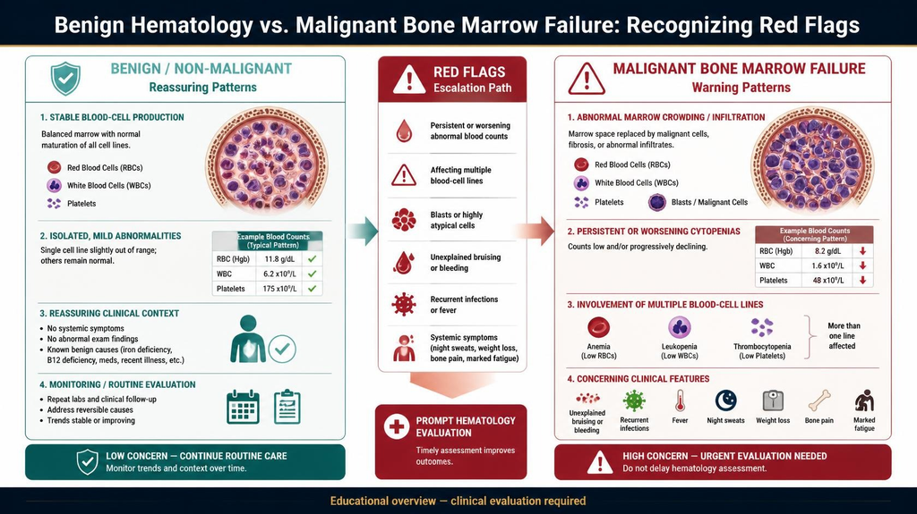
Not all hematologic conditions stem from malignancy.
The vast majority of blood abnormalities encountered in primary clinical practice are benign, reactive, or nutritional in origin.
However, the diagnostic challenge lies in distinguishing uncomplicated, reversible red blood cell deficits from progressive bone marrow failure syndromes such as Aplastic Anemia, Myelodysplastic Syndromes (MDS), or Myelofibrosis where the marrow’s stem cell microenvironment undergoes irreversible structural and genetic collapse.
Benign Hematology vs. Bone Marrow Failure Syndromes
| Clinical Category | Representative Conditions | Pathological Mechanism | Distinctive Clinical Presentation |
|---|---|---|---|
| Nutritional & Reactive Disorders | Iron deficiency, Vitamin B12 deficiency, Anemia of chronic inflammation. | Inadequate substrate availability or cytokine-induced iron sequestration; bone marrow architecture remains intact. | Isolated, gradual fatigue; responsive to targeted micronutrient repletion; normal leukocyte and platelet counts. |
| Hereditary Hemoglobinopathies | Thalassemia minor, Sickle Cell trait, Hereditary spherocytosis. | Inherited genetic mutations in globin gene synthesis or red cell membrane proteins causing premature destruction. | Lifelong mild anemia, microcytosis disproportionate to anemia degree, mild splenomegaly; stable across decades. |
| Autoimmune Cytopenias | Immune Thrombocytopenic Purpura (ITP), Autoimmune Hemolytic Anemia (AIHA). | Peripheral antibody-mediated destruction of mature circulating blood elements without primary stem cell failure. | Isolated thrombocytopenia (bruising) or sudden hemolytic jaundice; bone marrow shows hyperplastic compensatory response. |
| Primary Bone Marrow Failure | Aplastic Anemia, Myelodysplastic Syndromes (MDS), Pure Red Cell Aplasia. | Autoimmune stem cell destruction, clonal genetic mutations, or stromal fibrosis replacing hematopoiesis. | Pancytopenia (multi-lineage drop), progressive profound fatigue, spontaneous bleeding, and severe recurrent infections. |
Critical Blood Disorder Warning Signs That Demand Immediate Triage
While fatigue remains the common denominator across all blood conditions, certain companion manifestations termed “red flag” symptoms indicate that disease has spread beyond simple red cell loss:
- Multi-Lineage Cytopenias: When a standard blood panel demonstrates an unexplained drop in two or three cell lines simultaneously (anemia accompanied by absolute neutropenia or thrombocytopenia), primary bone marrow suppression must be evaluated immediately.
- Microvascular Bleeding & Petechiae: Spontaneous pinpoint reddish-purple spots on the lower limbs (petechiae), unprovoked hematomas, or bleeding gums signify a critical drop in functional platelets (< 20,000–30,000 / μL).
- Persistent Hepatosplenomegaly: Palpable enlargement of the spleen or liver suggests extramedullary hematopoiesis (the body attempting to produce blood outside the damaged marrow) or cellular infiltration.
- Refractory Anemia: Anemia that fails to improve despite therapeutic doses of iron, B12, or folate supplementation over a 4-to-6-week window.
Recognizing these key blood disorder warning signs early ensures patients avoid prolonged, ineffective oral therapies and receive prompt bone marrow evaluations.
Isolated microcytic anemia in a young adult is often nutritional. However, unexplained normocytic or macrocytic anemia in an individual over 50 especially when paired with borderline-low white cells or platelets warrants an immediate hematology consultation to rule out pre-leukemic myelodysplastic changes.
The Oncological Connection: How Blood Cancers Drive Deep, Unrelenting Fatigue
When fatigue stems from a hematologic malignancy, it behaves fundamentally differently from standard physiological tiredness. Known clinically as Cancer-Related Fatigue (CRF), this condition is characterized by a persistent, overwhelming sense of physical, emotional, and cognitive exhaustion that is disproportionate to recent activity and cannot be relieved by sleep or rest.
Understanding the specific signs of blood cancer fatigue helps patients and clinicians recognize when exhaustion is driven by malignant cellular processes rather than simple overwork or lifestyle stress.
The Biological Drivers of Malignant Fatigue
The profound energy depletion seen in blood cancers is caused by three interrelated pathological mechanisms:
- Bone Marrow Blast Overcrowding: In acute and chronic leukemias, rapidly dividing malignant blast cells overtake the marrow microenvironment. Healthy erythroid progenitor cells are physically and nutritionally displaced, causing severe, progressive normocytic anemia that starves peripheral muscles and vital organs of oxygen.
- Tumor Hypermetabolism (The Metabolic Drain): Malignant cells exhibit extreme rates of aerobic glycolysis (the Warburg effect). They consume massive quantities of systemic glucose, amino acids, and nutrients to fuel their uncontrolled division, effectively acting as a metabolic parasite that deprives healthy tissues of essential energy substrates.
- Chronic Inflammatory Cytokine Storm: Malignant lymphocytes, plasma cells, and the surrounding tumor microenvironment continuously secrete pro-inflammatory signaling proteins into the bloodstream notably Tumor Necrosis Factor-alpha (TNF-α), Interleukin-1 (IL-1), and Interleukin-6 (IL-6). These cytokines cross the blood-brain barrier, altering central neurotransmitter pathways, disrupting circadian rhythms, and triggering profound muscle catabolism.
How Fatigue Manifests Across Hematologic Subtypes
Different blood cancers combine this systemic energy drain with disease-specific physiological complications:
- Leukemias (AML / ALL / CML / CLL): Fatigue has a rapid, severe onset in acute forms, often accompanied by sudden pallor, low exercise tolerance, and dizziness due to precipitous drops in hemoglobin and platelet levels.
- Lymphomas (Hodgkin & Non-Hodgkin): Fatigue frequently co-occurs with systemic “B-symptoms,” including drenching night sweats, unprovoked low-grade fevers, and unintentional weight loss, driven by chronic lymphoid cytokine release.
- Multiple Myeloma: Fatigue is compounded by kidney strain (from light-chain protein accumulation) and hypercalcemia resulting from active osteolytic bone destruction, which causes severe muscle weakness, nausea, and cognitive clouding.
Fatigue Comparison: Lifestyle Burnout vs. Malignant Energy Drain
| Characteristic | Everyday Lifestyle Fatigue | Malignant / Blood Cancer Fatigue |
|---|---|---|
| Response to Rest | Improves significantly after 1–2 nights of restorative sleep or a restful weekend. | Persists unchanged despite prolonged bed rest or sleep; often worse upon waking. |
| Onset & Progression | Directly correlates with physical exertion, long working hours, or acute stress. | Arises without exertion; progressively worsens over weeks without an identifiable trigger. |
| Associated Physical Signs | Typically isolated; normal blood counts, no spontaneous bleeding or lymphadenopathy. | Accompanied by pallor, petechiae, unexplained bruising, persistent bone pain, or swollen nodes. |
| Impact on Daily Tasks | Causes mild sluggishness; basic daily routines remain manageable. | Severely limits basic self-care, walking short distances, or climbing a single flight of stairs. |
If severe, unremitting exhaustion is accompanied by unexplained weight loss, night sweats, recurrent fevers, or easy bruising, it should never be dismissed as stress. A thorough hematologic evaluation is essential to assess bone marrow health and rule out underlying malignancy.
The Diagnostic Workup: From Routine CBC to Bone Marrow Precision
Uncovering the root cause of chronic, unexplained exhaustion requires a structured, step-by-step diagnostic evaluation.
When investigating persistent exhaustion alongside unexplained cytopenias or circulating blast cells frequent early symptoms of leukemia fatigue a systematic diagnostic cascade ensures an accurate, definitive assessment, differentiating common nutritional deficiencies from complex bone marrow failure syndromes or hematologic malignancies.
Step-by-Step Hematologic Diagnostic Cascade
| Diagnostic Stage | Specific Tests Conducted | Primary Clinical Objective |
|---|---|---|
| Tier 1: Peripheral Blood Analysis | • Complete Blood Count (CBC) with differential• Manual Peripheral Blood Smear• Reticulocyte Count | Quantifies red cells, white cells, and platelets; identifies abnormal cellular morphology, poikilocytosis, or circulating immature blast cells. |
| Tier 2: Metabolic & Substrate Profiling | • Serum Ferritin, Iron, and Total Iron Binding Capacity (TIBC)• Vitamin B12 and Serum Folate levels• Liver (LFT), Kidney (KFT), and Serum LDH | Determines whether fatigue is driven by simple micronutrient depletion, chronic organ filtration strain, or high cellular turnover (elevated LDH/uric acid). |
| Tier 3: Bone Marrow Evaluation | • Bone Marrow Aspiration (cytological analysis)• Trephine Core Biopsy (histological architecture) | Evaluates marrow cellularity, structural architecture, fibrosis, and quantifies the exact percentage of immature malignant blast cells. |
| Tier 4: Molecular & Immunological Profiling | • Multiparametric Flow Cytometry (Immunophenotyping)• Cytogenetic Karyotyping & FISH panels• Next-Generation Sequencing (NGS) | Identifies lineage-specific surface markers (CD proteins), characteristic chromosomal translocations, and actionable driver mutations. |
Understanding the Key Diagnostic Milestones
- The Peripheral Blood Smear: While automated CBC analyzers flag numerical abnormalities, a manual peripheral smear examined under a microscope remains the primary visual gatekeeper. A hematopathologist checks for abnormal red cell shapes, hypersegmented neutrophils (signaling B12 deficiency), or the presence of immature myeloblasts or lymphoblasts.
- The Reticulocyte Production Index (RPI): Measures young red blood cells newly released from the marrow. A low reticulocyte count in an anemic patient confirms a production failure within the marrow, whereas a high reticulocyte count indicates peripheral destruction (hemolysis) or acute blood loss.
- Bone Marrow Aspiration and Biopsy: When multi-lineage cytopenias or blast cells are detected, a marrow examination at the posterior superior iliac crest provides definitive tissue confirmation. A blast count exceeding 20% in the marrow or peripheral blood establishes a diagnosis of acute leukemia according to WHO criteria.
When to Escalate to an Onco-Hematologist
Primary care physicians and patients should seek immediate hematology-oncology evaluation under the following clinical circumstances:
- Unexplained Bi-cytopenia or Pancytopenia: Concurrent reductions in red cells, white cells, or platelets without an obvious nutritional cause.
- Atypical Peripheral Cells: Smear reports indicating immature cells, leukoerythroblastic pictures, or suspected blast populations.
- Refractory Fatigue with Organomegaly: Persistent exhaustion paired with palpable enlargement of the spleen, liver, or lymph nodes.
Accurate hematologic diagnosis relies on complete immunophenotypic and cytogenetic profiling. Pinpointing the exact chromosomal abnormalities transforms a general diagnosis into a personalized, targeted treatment strategy.
Comprehensive Hematology & Oncology Care at Dasappa Cancer Hospital
Evaluating persistent, unexplained exhaustion requires clinical vigilance, rapid diagnostic turnaround, and access to dedicated oncological infrastructure. At Dasappa Cancer Hospital (Dasappa Memorial Institute of Oncology), our specialized clinical teams provide comprehensive diagnostic triage, detailed hematopathologic evaluations, and tailored systemic therapies in an empathetic, patient-first hospital setting.
Under the clinical guidance of Dr. Pandu Dasappa (M.Ch Surgical Oncology, GCRI Ahmedabad; FIAGES, FMAS; with over 27 years of surgical and clinical oncology experience), our multidisciplinary tumor boards collaborate with experienced medical oncologists and hematopathologists to ensure every patient receives an accurate diagnosis and an individualized, evidence-based management plan.
Key Clinical & Therapeutic Advantages at Dasappa Cancer Hospital
- Dedicated 75-Bed Super-Speciality Oncology Infrastructure: Purpose-built inpatient wards, intensive care units (ICU), and specialized isolation suites equipped to manage severe cytopenias, neutropenic fever, and acute hematologic complications.
- Specialized Day-Care Infusion & Transfusion Suites: Modern, comfortable day-care facilities staffed by certified oncology nursing teams for blood component transfusions (packed red cells, platelets) and administration of targeted therapies, monoclonal antibodies, and multi-agent chemotherapy regimens.
- Rapid Bone Marrow Diagnostics & Pathology: Streamlined processing for manual peripheral blood smears, bone marrow aspirations, trephine core biopsies, and molecular cytogenetic panels to eliminate critical diagnostic delays.
- Multidisciplinary Case Review: Complex hematologic and solid tumor presentations are reviewed by a collaborative board of surgical, medical, and diagnostic oncologists to ensure treatment protocols align with national and international guidelines (ICMR / NCCN).
Comprehensive patient counseling, dedicated oncology dietetics to manage cellular cachexia, and advanced palliative symptom support throughout the entire treatment continuum.
Conclusion
Persistent fatigue and weakness are not inevitable consequences of aging or busy urban schedules. When exhaustion persists despite rest, it serves as a vital biological indicator that warrants thorough medical investigation. Whether driven by treatable nutritional deficiencies, chronic organ strain, or underlying bone marrow failure, early diagnostic clarity is the key to effective intervention. Consulting a specialized oncology team ensures that serious hematologic conditions are caught early, protecting vital organ health and restoring long-term vitality.
Schedule a Clinical Evaluation at Dasappa Cancer Hospital
If you or a loved one is experiencing persistent exhaustion, unprovoked bruising, recurring infections, or abnormal routine blood counts, contact our clinical oncology team in Bengaluru for an expert assessment:
- Hospital Address:
Dasappa Memorial Institute of Oncology (Dasappa Cancer Hospital),
No. 132, Gandhi Bazaar Main Road, Basavanagudi, Bengaluru, Karnataka – 560004 - Consultation Helpline: +91 80 2667 9294 / +91 98450 12345
- Website: dasappacancerhospital.com
Specialties Available: Medical Oncology, Surgical Oncology, Day-Care Chemotherapy, Hematology Diagnostics, Second Opinion Consultations.