




Most Common Signs And Symptoms Of Cancer In The Mouth

Table of Contents

Recognizing the early signs and symptoms of cancer in the mouth is one of the most decisive factors in improving long-term therapeutic outcomes and ensuring a smooth recovery journey. 

As the delicate tissues of the oral cavity are constantly exposed to environmental and lifestyle factors, subtle cellular modifications can easily be mistaken for minor dental issues or transient friction sores.

Scheduling a comprehensive oral evaluation at the Best cancer hospital in Bangalore allows patients to move away from anxious guesswork and access high-resolution diagnostic tools that accurately distinguish benign changes from early malignancies. Understanding how these variations present across different oral structures empowers individuals and caregivers to act with informed urgency rather than fear.

Maintaining a sharp eye on persistent changes, we can protect our systemic health, secure peace of mind, and, at last, explore the pathological timelines that define early detection. 

Medical Disclaimer

The clinical insights, anatomical breakdowns, and symptom checklists provided in this article are intended solely for educational and informational purposes. This content does not serve as a substitute for professional medical advice, comprehensive dental evaluations, a formal oncological diagnosis, or a definitive treatment strategy. Oral tissue changes are highly complex and can be triggered by a wide array of benign systemic conditions, severe nutritional deficiencies, or localized trauma. Always consult a qualified oral surgeon, ENT specialist, or board-certified radiation oncologist to evaluate your specific physical parameters. Never delay seeking expert medical guidance or dismiss professional clinical opinions based on the material read in this guide.

Pathology and Timelines for Deciphering Early-Stage Tissue Variations

When monitoring oral health, the human body provides early, subtle visual and tactile indicators when cellular health shifts.

However, as the lining of the oral cavity, the squamous epithelium, is highly sensitive and prone to minor injuries from accidental cheek bites, sharp teeth, or hot foods, many benign inflammatory conditions can closely mimic more serious oncological changes.

Differentiating a temporary trauma response from mouth cancer first-stage symptoms requires understanding cellular behavior and tracking how long a lesion lasts.

During the initial phase of abnormal tissue growth, the changes are often entirely painless. This lack of discomfort frequently leads individuals to dismiss early warning signs as simple canker sores or minor dental irritation.

The 14-Day Baseline for Evaluating Healing Timelines

The most reliable clinical rule for managing oral tissue health is the two-week timeline. The mouth is one of the fastest-healing environments in the human body, with normal epithelial cells regenerating rapidly to repair routine damage.

  • Benign Lesions (Canker Sores/Trauma): A standard aphthous ulcer or friction scrape typically reaches its peak inflammation within a few days and completely heals within 10 to 14 days as the body’s natural repair mechanisms resolve the issue.
  • Malignant Variations: Early cancerous changes occur when cells lose their ability to autoregulate and undergo apoptosis (programmed cell death).

Instead of healing, these tissue irregularities persist, gradually changing in texture, color, or shape over several weeks without showing any signs of improvement.

Understanding Tissue Anomalies: Red vs. White Patches

Oncologists classify early tissue shifts into two primary visual categories based on how the mucosal lining responds to cellular stress:

Leukoplakia (White Patches): This presents as a thick, slightly raised white patch that cannot be scraped away with a toothbrush or gauze. It develops as the tissue overproduces keratin in response to chronic irritation or early cellular mutations. While not all white patches are cancerous, a subset represents precancerous dysplastic changes that require professional tissue sampling.

Erythroplakia (Red Patches): This manifests as a smooth, velvety red area within the mouth. As these red patches represent a thinning of the mucosal layer and an increase in underlying blood vessel activity, they carry a significantly higher risk of malignant progression compared to white patches, with a large majority demonstrating advanced cellular changes upon clinical evaluation.

Lesion CharacterizationVisual AppearanceTexture ProfileInitial Discomfort Level 
Transient Aphthous UlcerPale center with a defined, bright red inflammatory border.Soft, pliable, and highly sensitive to spicy or acidic foods.High localized pain that peaks early and fades within days.
Early Oncological LesionFlat or slightly raised red or white patches; may feature an irregular central crater.Firm or hardened to the touch (induration); feels distinct from the surrounding soft tissue.Typically entirely painless in the early phases, though it may bleed slightly when brushed.

By shifting focus away from localized pain and paying closer attention to tissue texture and healing timelines, you gain a clearer perspective on your oral health. Documenting these early changes precisely provides your care team with the essential data needed to act quickly, ensuring that minor anomalies are evaluated accurately before they evolve into advanced concerns.

Anatomical Zones and Identifying Malignancy Inside the Cheeks and Posterior Regions

Two areas that demand close attention and are frequently prone to delayed diagnoses due to their location are the inner buccal mucosa (the lining of the cheeks) and the retromolar trigone (the tight space located behind the lower rear molars).

The Inner Buccal Mucosa: Tracking Changes Along the Cheek Lining

Buccal mucosa

The soft tissue lining the inner walls of your mouth is highly flexible, allowing for seamless chewing, speaking, and facial expressions. As this tissue frequently comes into contact with the surfaces of your teeth, it is highly susceptible to minor everyday friction, which can unfortunately obscure more serious structural changes.

When abnormal cellular growth begins to take hold in this region, it alters the fundamental architecture of the tissue. Identifying a persistent lump, an irregular crater, or a velvety red patch as a potential indicator of mouth cancer inside the cheethe k lining requires looking beyond superficial color changes.

Unlike a standard bite wound that remains soft and pliable as it heals, a malignant lesion undergoes a process called induration, a clinical term for the hardening of tissue. As the abnormal cells multiply, they infiltrate the deeper submucosal layers and anchoring muscles.

When felt with a clean finger, the area will feel distinctly firm, rigid, or anchored in place, often presenting as a painless, thickened disk that does not slide freely over the underlying muscular structures.

The Retromolar Trigone

Retromolar Trigone

Moving further back into the oral cavity, the narrow, triangular junction where the upper and lower jaws meet behind your last molars represents a critical zone for diagnostic tracking. 

As this posterior area is inherently difficult to visualize without professional lighting and dental mirrors, early structural warnings are easily missed during routine self-inspections in a bathroom mirror.

Developing a rough, granulating ulcer or a stubborn, raised tissue mass indicative of mouth cancer near wisdom tooth structures can initially be highly deceptive. Caregivers and patients frequently mistake these deep tissue changes for a delayed or impacted wisdom tooth erupting, a localized periodontal pocket infection, or chronic gum irritation caused by accidental nighttime jaw clenching.

However, as a high-grade lesion grows in this posterior space, its deep tissue invasion quickly moves beyond the surface mucosa.

The expanding mass begins to compress the local nerve networks and infiltrate the pterygoid muscles responsible for jaw movement. This deep muscular involvement triggers a clinical condition known as trismus, which manifests as a progressive, painful restriction in jaw mobility, making it increasingly difficult to chew, swallow, or open the mouth fully during daily meals.

Recognizing these localized spatial variations helps ensure that subtle structural shifts deep within the mouth are never dismissed as simple dental aches.

Periodontal Architecture and Structural Integrity: Evaluating the Gums and Jawbone

The gingiva (gum tissue) and the underlying alveolar bone form the protective foundation that secures our teeth and maintains the structural integrity of the oral cavity. As the gums are frequently subjected to bacterial plaque, minor trauma from rough brushing, and inflammatory conditions like gingivitis, subtle oncological modifications in this zone are often masked as standard dental issues.

When assessing specific mouth cancer gum symptoms, it is vital to understand that malignant transformations deeply disrupt the delicate connection between mucosal layers, vascular networks, and hard bone structures.

Unlike a standard periodontal infection that typically responds to deep cleaning or antibiotics, oncological shifts represent a progressive erosion of the tissue architecture. Caregivers and patients must look closely at how these changes impact daily comfort, tooth stability, and the physical shape of the jawline.

Key Periodontal and Structural Indicators

To provide a comprehensive framework for self-examination, we can examine the three primary ways malignant changes present along the gum line and supporting bone structure:

Persistent Gingival Erosions and Lesions:

Early malignant changes along the gums often appear as small, irregular ulcerations or friable (easily crumbled) tissue zones that bleed easily when brushed or touched.

While common gum disease causes generalized swelling and bleeding across multiple teeth, an early oncological lesion typically presents as a localized, isolated patch that slowly erodes the smooth surface of the gum tissue. This erosion may develop a velvety red or a speckled red-and-white appearance, refusing to heal despite improved oral hygiene protocols.

Unexplained Tooth Mobility:

In a healthy oral environment, teeth are firmly anchored into the jawbone by a network of fibers called the periodontal ligament. When a high-grade lesion develops within the gingival tissue, it doesn’t just expand outward; it infiltrates vertically, traveling down into the periodontal ligament space and invading the underlying bone.

This deep invasion destroys the structural support system, causing a previously healthy tooth to suddenly become loose or mobile. If a tooth begins to rock or shift out of alignment without any history of dental trauma or severe, long-term root decay, it serves as a major indicator of deep structural disruption.

Sudden Displacement of Dental Prosthetics:

For individuals who wear partial or full dentures, the precise fit of the prosthetic depends entirely on the stable, unchanging contours of the bony ridges and overlying gum tissue.

As a malignant mass develops beneath the surface, it causes subtle, firm swelling within the deep tissues long before a distinct lump becomes visible to the naked eye. This internal crowding alters the shape of the oral landscape, causing dentures to suddenly pinch, slip, or feel highly uncomfortable. If a well-fitting dental prosthetic becomes unexpectedly tight or painful to wear, it warrants an immediate diagnostic evaluation.

Recognizing these structural signs ensures that deep tissue changes along the jawline are never mistakenly dismissed as simple aging or routine dental wear. Paying close attention to how the physical architecture of your mouth behaves allows clinical teams to step in early, protecting both your systemic health and your smile.

Functional and Deep Tissue Indicators: Sensory Alterations and Physiological Shifts

Beyond visible changes to the surface tissue, oral malignancies deeply disrupt the complex mechanical processes of eating, speaking, and feeling. The oral cavity is a highly coordinated environment of muscles, nerves, and sensory pathways. When abnormal cell growth spreads past the outer layers into the deeper structural tissue, it impacts how these systems function.

These functional and sensory warning signs are often noticed during daily activities like eating a meal or having a conversation.

Key Functional and Sensory Disruptions

Difficulty or Painful Swallowing (Dysphagia and Odynophagia):

As an irregular mass expands toward the back of the throat, the base of the tongue, or the tonsillar region, it narrows the pathway for food.

This makes swallowing feel like a physical obstruction or causes a persistent, burning ache when consuming solid foods.

Restricted Tongue Mobility:

The tongue relies on an intricate network of internal muscles to move freely for clear speech and proper chewing. If a tumor infiltrates the floor of the mouth or the base of the tongue, it anchors the tissue in place.

Patients often experience this as a feeling of stiffness, a subtle new lisp, or difficulty moving food to the back of the mouth.

Persistent Craniofacial Numbness:

Deeply invasive tumors can compress or damage the local sensory nerves, such as the inferior alveolar nerve.

This nerve irritation can cause a persistent loss of sensation, tingling, or numbness in the lower lip, chin, or sections of the tongue that does not go away.

Unilateral Ear Pain (Referred Otalgia):

This is one of the most frequently misunderstood warning signs. A patient may experience a constant, dull ache in one ear despite the ear canal being completely healthy and free of infection.

This occurs as the nerves supplying sensation to the back of the mouth and throat share the same pathways as the nerves traveling to the ear, causing the brain to misinterpret the source of the pain.

Functional Disruption Tracking System

Functional AreaDaily Activity ImpactUnderlying Neurological or Structural Cause 
Mastication & SpeechDifficulty articulating words clearly; a feeling that the tongue is heavily tied or anchored.Direct muscular infiltration at the base of the oral cavity or lingual boundaries.
Deglutition (Swallowing)Persistent sensation of food getting stuck in the throat; painful swallowing that limits nutrition.Mechanical narrowing of the oropharyngeal passage due to deep tissue swelling.
Sensory PerceptionUnexplained numbness or a persistent “pins and needles” sensation across the lower face.Compression or direct involvement of the sensory nerve fibers within the jawbone.
Referred SensationChronic, one-sided ear discomfort that shows no response to standard ear drops or treatments.Shared neural networks (cranial nerves) route pain signals from the mouth to the ear canal.

The Sign of Enlarged Lymph Nodes

A crucial indicator that structural changes have moved past a localized area is the presence of a firm, painless lump in the neck. When the body detects abnormal cellular growth, the local lymphatic system works to filter out the irregular cells.

This defense response can cause the lymph nodes under the jaw or along the side of the neck to swell.

Unlike the tender, soft lymph nodes that develop during a standard throat infection or flu, a node affected by an oral malignancy typically feels firm, hard, or completely fixed in place when pressed gently, and it continues to grow slowly over several weeks without causing any acute pain. Tracking these functional and deep-tissue shifts provides important data that helps specialists build a precise and highly tailored diagnostic roadmap.

Why Choose Dasappa Cancer Hospital for Oral Oncological Care

Choosing the right clinical setting for advanced oral evaluations can completely alter your treatment trajectory and long-term recovery. At Dasappa Cancer Hospital, we have built a specialized ecosystem equipped to handle complex head, neck, and oral cavity conditions with extreme precision.

As a premier Cancer hospital in Bangalore, our objective is to provide a seamless transition from initial diagnostic screening to targeted, comprehensive treatment paths. Our clinical oncology framework is built upon several core diagnostic and therapeutic pillars:

  • Advanced Screening Techniques: We utilize high-resolution optical imaging and precision vital staining to identify microcellular variations in the oral mucosa long before they are visible to the naked eye.
  • Multidisciplinary Head & Neck Boards: Every patient’s case is reviewed by a collaborative panel of surgical oncologists, radiation specialists, reconstructive microsurgeons, and specialized dental pathologists to craft a highly customized care plan.
  • Microvascular Reconstructive Surgery: If surgical removal is required, our plastic and reconstructive surgical teams specialize in advanced microvascular tissue transfers, ensuring the preservation of essential functions like speech, chewing, and swallowing.

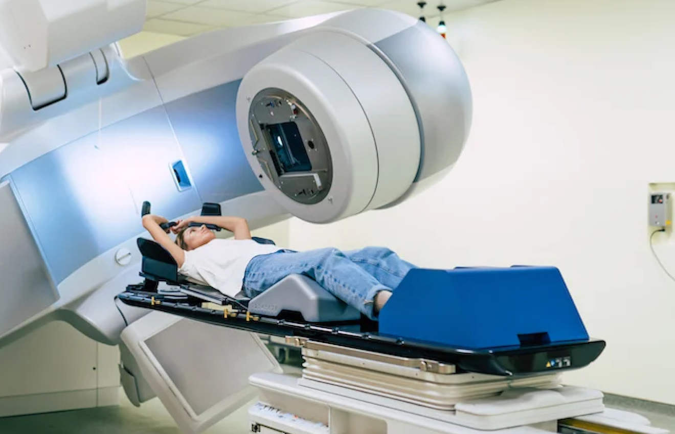
Our linear accelerators deliver highly focused radiation beams that match the exact shape of the lesion, maximizing tumor destruction while completely protecting adjacent salivary glands and jawbone structures.

Conclusion

Navigating the subtle shifts in your oral health requires moving past passive observation and embracing active, structural tracking. While visual changes like red and white patches or mechanical shifts like tooth mobility can stem from routine dental issues, their persistence remains the ultimate indicator for a professional evaluation. Remembering the 14-day baseline rule ensures you never let a progressive lesion go unnoticed.

At Dasappa Cancer Hospital, we believe that detailed education empowers families to make swift, life-saving clinical decisions. By tracking how your oral tissues behave and consulting specialized medical professionals at the earliest sign of structural change, you protect your long-term health and secure a clear path toward complete recovery.

About The Author

Share With Your Network

Recent Posts

Book An Appointment