




10 Common Symptoms Of Glioblastoma

Table of Contents

Navigating a brain health diagnosis can be daunting, especially when the warning signs are subtle or masked by everyday stressors.

Recognizing the early glioblastoma symptoms is a critical step in facilitating timely medical intervention and personalized care. At the Best cancer hospital in Bangalore, we emphasize that early identification of neurological changes provides the greatest opportunity for effective therapeutic planning. While these indicators often overlap with less severe conditions, persistent neurological shifts demand a professional evaluation to ensure structural health.

Understanding these markers empowers patients and families to move beyond uncertainty, bridging the gap between subtle behavioral changes and expert medical assessment. Prioritizing clarity and diagnostic precision, we support individuals in navigating the complexities of neuro-oncology with informed confidence.

Medical Disclaimer

The educational concepts, symptom breakdowns, and clinical information shared in this article are provided strictly for informational purposes. This content does not substitute for professional medical advice, a formal diagnosis, or a definitive clinical treatment plan. Neurological presentations are highly complex and can be caused by a wide variety of medical conditions; only a qualified neurologist or neuro-oncologist can accurately assess your individual health parameters. Never ignore or delay seeking professional medical consultation based on the information provided here. If you are experiencing an acute neurological event, such as a seizure or sudden loss of motor function, please seek emergency medical attention immediately.

Defining the Pathology of Understanding Glioblastoma

To truly grasp the clinical implications and the reason behind the onset of various neurological signs, one must first ask: What is GBM?

Glioblastoma Multiforme (GBM) is classified as a Grade IV astrocytoma, the most aggressive and highly invasive form of primary brain cancer. Unlike tumors that start elsewhere in the body and metastasize to the brain, GBM originates within the glial cells, which are the support staff of your nervous system. 

These cells, specifically astrocytes, are responsible for maintaining the blood-brain barrier and providing nutrients to neurons. In a glioblastoma, these cells lose their regulatory control, multiplying rapidly and creating a chaotic, poorly organized mass of malignant tissue.

The Mechanism of Malignancy and How the Tumor Operates

The symptoms patients experience are rarely just the result of the tumor mass itself. They are a combination of direct tissue invasion and the mass effect, the pressure the tumor exerts on the surrounding healthy brain. Glioblastomas are infamous for their finger-like projections that infiltrate the surrounding brain architecture, making them notoriously difficult to remove entirely with surgical intervention alone.

To visualize how this pathology affects the cranial environment, consider the following clinical breakdown:

Pathological FeatureScientific DefinitionImpact on Neurological Function 
Cellular HeterogeneityThe tumor contains many different types of cells with varying genetic mutations.Makes the malignancy resistant to standardized, uniform therapy.
AngiogenesisThe tumor rapidly builds its own dedicated, leaky blood vessels.Causes fluid accumulation (edema) and increased intracranial pressure.
Infiltrative GrowthInvasive tendrils extend deep into healthy, functional white matter.Disrupts the communication pathways between different brain regions.
Necrotic CoreCentral areas of the tumor die due to rapid, unsustainable growth.Triggers inflammatory responses that further exacerbate brain swelling.

The Cranial Crowding Effect: A Conceptual Map

Imagine the human brain as a highly organized city contained within a rigid, immovable wall, the skull.

As the skull cannot expand to accommodate new growth, the presence of an expanding, fluid-leaking mass (the tumor and its associated edema) creates a dangerous crowding effect.

  • The Displacement Factor: As the tumor grows, it literally pushes aside healthy neurons. If it pushes on the area controlling speech, speech is lost. If it pushes on the area controlling movement, paralysis occurs.
  • The Pressure Gradient: The increase in internal cranial pressure creates a secondary cascade of issues. This is why many patients do not feel sick in the traditional sense initially; they experience mechanical problems, such as morning headaches caused by the shifting of fluid pressure while lying flat.

This distinction is vital for patients and caregivers: Glioblastoma is not just a disease of cell growth; it is a disease of space and displacement.

Recognizing that your symptoms are the result of the brain responding to this internal pressure helps demystify why neurological markers can appear suddenly or fluctuate in severity.

Understanding this biology is the first step in our multidisciplinary approach at Dasappa Cancer Hospital, where we focus not just on tumor suppression but on managing the systemic environment of the brain to preserve your quality of life.

Cognitive and Behavioral Shifts are the Early Indicators

The brain functions as the command center for every thought, emotion, and action. As the frontal and temporal lobes are large, active areas of the brain, they are frequently the sites where high-grade lesions manifest.

When a mass begins to expand or infiltrate these regions, the earliest indicators often appear not as physical pain, but as subtle, persistent shifts in a person’s cognitive baseline and personality.

Recognizing these changes requires observing the individual’s normal behavioral baseline compared to their current state.

1. Persistent or Worsening Headaches

Headache

While many people experience tension headaches, those associated with intracranial pressure are distinct. Patients often report that their discomfort is most intense in the early morning. This occurs as, while lying flat during sleep, the venous drainage from the brain is slightly reduced, causing intracranial pressure to spike. As the day progresses and the patient sits up, the pressure can shift, potentially causing the intensity of the pain to fluctuate.

2. Personality and Behavioral Changes

Behavioral Changes

The frontal lobe of the brain governs social inhibition, decision-making, and emotional regulation. If a mass disrupts this specific architecture, friends and family may notice uncharacteristic behavioral shifts:

  • Reduced Inhibition: Engaging in socially inappropriate behaviors or saying things out of character.
  • Apathy and Withdrawal: A sudden loss of interest in hobbies, career, or personal relationships.

Emotional Lability and Rapid, extreme mood swings, such as moving from calm to intense irritability without an obvious external trigger.

3. Cognitive Decline and Memory Loss

Memory loss

Unlike standard age-related forgetfulness, which tends to be gradual and consistent, cognitive shifts associated with a mass effect often feel foggy and rapidly progressive. Patients may report:

  • Focus Deficits: An inability to maintain concentration on a single task, such as reading a newspaper or following a simple conversation.
  • Word-Finding Difficulties: Struggling to recall common nouns or names of close friends.

Confusion and Occasional lapses in spatial orientation, such as becoming disoriented in familiar environments.

The Behavioral Audit Table

For caregivers and family members, it can be difficult to quantify these changes. The following table provides a way to categorize these early cognitive markers for professional reporting:

Observation CategoryNormal BaselinePotential Red Flag 
Social RegulationExercises appropriate social filters.Displays sudden, aggressive, or uncharacteristic social outbursts.
Decision MakingCapable of routine tasks (paying bills, scheduling).Shows impulsive decision-making or inability to complete simple, logical steps.
AlertnessConsistent focus during waking hours.Frequent zoning out or uncharacteristic apathy toward surroundings.

Why Cognitive Markers Matter?

These cognitive markers are often the first to be dismissed as stress or fatigue, but they serve as essential diagnostic data. If these behavioral shifts persist or show a pattern of worsening over several weeks, they should be documented clearly and discussed during your next neurological consultation to help clinical teams piece together the full picture of the patient’s neurological state.

Motor and Physical Disruption of Clinical Manifestations of Neural Interference

While cognitive shifts are often the most subtle and early indicators of a high-grade intracranial lesion, motor and physical manifestations are typically the result of direct structural invasion or compression of the motor cortex, cerebellum, or descending nerve pathways. When identifying potential symptoms of brain cancer, it is crucial to recognize that physical deficits often present unilaterally, meaning they affect only one side of the body.

This pattern occurs as the human brain is cross-wired; a lesion in the left hemisphere typically manifests as weakness or coordination failure on the right side of the body.

4. Seizures

Seizure

For many patients, a sudden, unexpected seizure is the very first, definitive clinical sign that something is amiss. A brain tumor acts as an irritable focus within the neural architecture. The rapid, uncontrolled proliferation of malignant cells can disrupt the normal electrical firing patterns of neurons, leading to sudden, rhythmic, and involuntary electrical discharges. These can range from focal seizures, where a patient might experience twitching in just a hand or a facial muscle, to generalized seizures that result in a loss of consciousness and full-body convulsions.

5. Motor Weakness (Hemiparesis)

Weakness

Motor weakness, or hemiparesis, is the gradual or sudden loss of voluntary muscle control. This typically involves the arm, leg, or facial muscles on one side of the body. As the mass displaces the motor cortex, the pathways that send movement signals from the brain to the muscles become blocked or distorted. Families often notice this during routine tasks:

  • Grip Strength: Difficulty opening a jar or holding a pen.
  • Gait Asymmetry: A subtle dragging of one foot or a limp when walking.

Facial Droop and A slight, asymmetrical lowering of one corner of the mouth or eyelid.

6. Balance and Coordination Issues (Ataxia)

Balancing issue

If a mass develops near the cerebellum (the back of the brain) or the brainstem, the patient’s ability to coordinate fine motor movements is often compromised. Ataxia refers to a lack of muscle control or coordination of voluntary movements. It is not necessarily muscle weakness, but rather a disruption in the data processing required to walk, stand, or reach for objects.

Diagnostic Triage: Categorizing Motor Deficits

To assist our clinical teams in the triage process, we categorize motor physical deficits based on the specific neural pathways they likely involve. The table below serves as a professional guideline for identifying when these physical shifts require an immediate neurological investigation:

Symptom CategoryUnderlying Anatomical InvolvementObserved Clinical Manifestation 
Electrical IrritabilityMotor Cortex / Cortical surfaceFocal twitching, involuntary shaking, loss of consciousness (seizure).
Voluntary Motor DeficitMotor Cortex / Descending tractsProgressive weakness in an arm or leg, difficulty lifting objects.
Coordination DeficitCerebellum / BrainstemWide-based gait, drunken walk, inability to touch finger to nose.
Reflex DysfunctionSpinal tracts / BrainstemHyper-reflexive jerks or sudden, spastic muscle tone changes.

Why does Physical Asymmetry Matters?

It is essential to understand that these motor deficits are rarely fixed. They often fluctuate. A patient might wake up with severe arm weakness that improves slightly by the afternoon as fluid pressure (edema) shifts.

Clinical Observation: When assessing these symptoms, document whether the weakness is transient (comes and goes) or progressive (gets consistently worse). This distinction is vital.

A progressive motor deficit is a clear, urgent indicator that the tumor is actively displacing or damaging the neural architecture. If you or a loved one experiences any of these physical warning signs, do not wait for them to resolve on their own; these are significant neurological events that require the rapid diagnostic expertise available at a specialized neuro-oncology centre.

Neurological, Sensory, and Systemic Indicators

As the mass effect of malignant tumor brain progression continues to exert pressure on the surrounding cerebral architecture, clinical indicators often move beyond simple motor or cognitive deficits. 

These later-stage indicators involve the intricate sensory processing pathways and physiological regulation systems. As the brain serves as the central hub for sensory interpretation, integrating input from our eyes, ears, and internal receptors, any disruption in these regions manifests as profound changes in perception and daily function.

7. Nausea and Vomiting

Nausea and vomiting

Nausea and vomiting are classic signs of increased intracranial pressure (ICP). This often occurs without the presence of traditional gastrointestinal symptoms like food poisoning or viral illness.

Patients frequently report that this nausea is most severe in the morning, often subsiding after a period of being upright. This occurs as the recumbent position during sleep allows intracranial pressure to build, stimulating the vomiting center located in the medulla oblongata within the brainstem.

8. Vision Changes

Vision change

Vision is a complex process involving not just the eyes, but the optic nerves and the visual cortex at the back of the brain (the occipital lobe). If the tumor invades or compresses the optic pathways, patients may report persistent blurring, double vision (diplopia), or a loss of peripheral field, often described as bumping into door frames or missing objects on one side of their plate.

These Sensory symptoms are frequently ignored until they significantly interfere with driving or reading, but they are critical indicators of direct neural path interference.

9. Speech Difficulty (Aphasia)

Speech difficulty

Language processing is primarily handled by the dominant hemisphere (typically the left). A lesion in Broca’s or Wernicke’s areas of the brain can disrupt the ability to translate thoughts into articulate speech. This is known as aphasia. It may manifest as:

  • Expressive Aphasia: The patient knows exactly what they want to say but cannot physically form the words.
  • Receptive Aphasia: The patient can speak clearly, but the words they choose are nonsensical or do not match the context of the conversation.

10. Chronic Fatigue

Fatigue

While being tired is common, the chronic lethargy associated with brain cancer is systemic and profound. It is not solved by sleep. This fatigue arises from the body’s massive expenditure of energy trying to compensate for the tumor’s impact on metabolic regulation and the sheer exhaustion of the brain managing its own internal displacement.

It feels less like a lack of sleep and more like an overwhelming, persistent heaviness that makes even the simplest daily activities feel insurmountable.

Triage Chart: Identifying Sensory and Physiological Red Flags

The following chart outlines these indicators to help patients and families differentiate between transient fatigue and serious neurological red flags:

SymptomPrimary Physiological CauseUrgent Clinical Indicator 
NauseaElevated Intracranial Pressure (ICP)Vomiting without nausea or gastric distress.
Vision LossOptic Nerve/Pathway CompressionSudden loss of peripheral or central visual fields.
Speech LossDamage to language centers (Aphasia)Inability to form coherent sentences or comprehend speech.
Deep FatigueSystemic Metabolic StrainPersistent lethargy unresponsive to rest.

Why does recording symptoms matter?

Recognizing these symptoms is not about creating alarm; it is about establishing a clear, documented record of neurological change.

These indicators represent the brain’s struggle to maintain function in the face of abnormal internal growth. Documenting these specific changes, how often they occur, whether they are worsening, and the context in which they happen provides our specialists with the essential, high-resolution data needed to begin an effective neuro-oncological management plan.

Why Choose Dasappa Cancer Hospital for Neuro-Oncological Care?

When facing a diagnosis involving high-grade lesions, the experience and infrastructure of your medical team are the most significant factors in determining your care trajectory. At Dasappa Cancer Hospital, we specialize in comprehensive brain tumor treatment in Bangalore, integrating cutting-edge surgical precision with advanced neuro-radiology and medical oncology to offer a unified, patient-centric approach.

We understand that a diagnosis of glioblastoma requires more than just standard medical intervention; it demands a dedicated, multidisciplinary environment where every decision is driven by the latest clinical data. Our commitment to your care includes:

  • Multidisciplinary Neuro-Oncology Boards: Our treatment plans are formulated by a team of neurosurgeons, radiation oncologists, and medical oncologists who collaborate to tailor your protocol based on the specific genetic and structural markers of your condition.
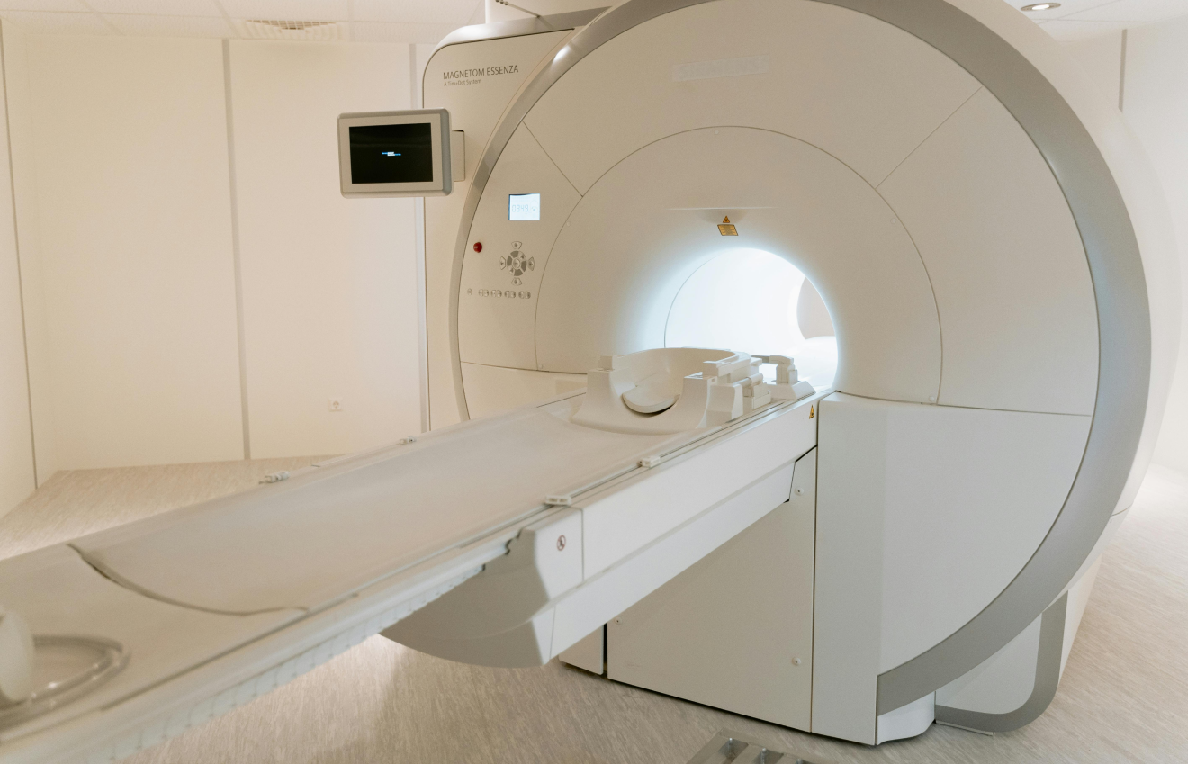
  • Precision Imaging & Guidance: We utilize advanced neuro-imaging suites to map the tumor’s infiltration, ensuring that surgical and radiation planning is performed with microscopic accuracy to maximize resection and minimize damage to critical neural pathways.
  • Comprehensive Symptom Management: Beyond tumor destruction, our palliative and supportive care teams focus on mitigating side effects such as cerebral edema (swelling) and seizures, ensuring your physical comfort and cognitive stability throughout the treatment duration.

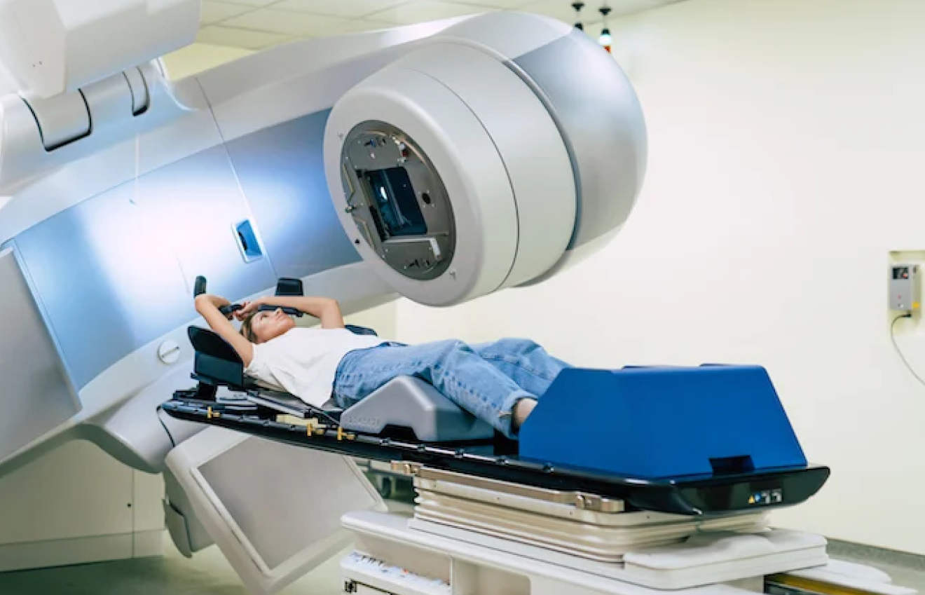
From targeted radiotherapy protocols to the latest in chemotherapy options, we provide access to evidence-based therapies that are specifically geared toward the aggressive nature of high-grade glial malignancies.

Conclusion

Recognizing the subtle shifts in your neurological baseline is the most critical first step toward effective management and long-term care. While the symptoms discussed, ranging from cognitive and behavioral changes to motor and sensory deficits, can be alarming, they are vital pieces of information that guide our clinical teams toward the right diagnostic and therapeutic pathway.

At Dasappa Cancer Hospital, we view every patient as a partner in their own care journey. Our mission is to move you from the uncertainty of symptoms to the clarity of a structured, expert-led management plan. If you or a loved one is experiencing persistent neurological shifts, do not delay a professional consultation. Early detection remains the most effective tool we have to preserve brain function and maintain your quality of life. 

About The Author

Share With Your Network

Recent Posts

Book An Appointment